Elham Rahimian, Felice D'Arco, Sniya Sudhakar, Majid R Tahsini, Neda Azin, Mahdis Morovvati, Parvaneh Karimzadeh, Mohammad Aidin Farahvash
{"title":"The full spectrum of MRI findings in 18 patients with Canavan disease: new insights into the areas of selective susceptibility.","authors":"Elham Rahimian, Felice D'Arco, Sniya Sudhakar, Majid R Tahsini, Neda Azin, Mahdis Morovvati, Parvaneh Karimzadeh, Mohammad Aidin Farahvash","doi":"10.1007/s00234-024-03388-x","DOIUrl":null,"url":null,"abstract":"<p><strong>Introduction: </strong>Canavan disease (CD) is a rare autosomal recessive neurodegenerative disorder caused by a deficiency of aspartoacylase A, an enzyme that degrades N-acetylaspartate (NAA). The disease is characterized by progressive white matter degeneration, leading to intellectual disability, seizures, and death. This retrospective study aims to describe the full spectrum of magnetic resonance imaging (MRI) findings in a large case series of CD patients.</p><p><strong>Materials and methods: </strong>MRI findings in 18 patients with confirmed CD were investigated, and the full spectrum of brain abnormalities was compared with the existing literature to provide new insights regarding the brain MRI findings in these patients. All the cases were proven based on genetic study or NAA evaluation in urine or brain.</p><p><strong>Results: </strong>Imaging analysis showed involvement of the deep and subcortical white matter as well as the globus pallidus in all cases, with sparing of the putamen, caudate, and claustrum. The study provides updates on the imaging characteristics of CD and validates some underreported findings such as the involvement of the lateral thalamus with sparing of the pulvinar, involvement of the internal capsules and corpus callosum, and cystic formation during disease progression.</p><p><strong>Conclusion: </strong>To our knowledge, this is one of the largest case series of patients with CD which includes a detailed description of the brain MRI findings. The study confirmed many of the previously reported MRI findings but also identified abnormalities that were previously rarely or not described. We speculate that areas of ongoing myelination are particularly vulnerable to changes in CD.</p>","PeriodicalId":19422,"journal":{"name":"Neuroradiology","volume":" ","pages":"1829-1835"},"PeriodicalIF":2.4000,"publicationDate":"2024-10-01","publicationTypes":"Journal Article","fieldsOfStudy":null,"isOpenAccess":false,"openAccessPdf":"","citationCount":"0","resultStr":null,"platform":"Semanticscholar","paperid":null,"PeriodicalName":"Neuroradiology","FirstCategoryId":"3","ListUrlMain":"https://doi.org/10.1007/s00234-024-03388-x","RegionNum":3,"RegionCategory":"医学","ArticlePicture":[],"TitleCN":null,"AbstractTextCN":null,"PMCID":null,"EPubDate":"2024/6/17 0:00:00","PubModel":"Epub","JCR":"Q2","JCRName":"CLINICAL NEUROLOGY","Score":null,"Total":0}
引用次数: 0
Abstract
Introduction: Canavan disease (CD) is a rare autosomal recessive neurodegenerative disorder caused by a deficiency of aspartoacylase A, an enzyme that degrades N-acetylaspartate (NAA). The disease is characterized by progressive white matter degeneration, leading to intellectual disability, seizures, and death. This retrospective study aims to describe the full spectrum of magnetic resonance imaging (MRI) findings in a large case series of CD patients.
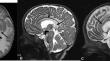
Materials and methods: MRI findings in 18 patients with confirmed CD were investigated, and the full spectrum of brain abnormalities was compared with the existing literature to provide new insights regarding the brain MRI findings in these patients. All the cases were proven based on genetic study or NAA evaluation in urine or brain.
Results: Imaging analysis showed involvement of the deep and subcortical white matter as well as the globus pallidus in all cases, with sparing of the putamen, caudate, and claustrum. The study provides updates on the imaging characteristics of CD and validates some underreported findings such as the involvement of the lateral thalamus with sparing of the pulvinar, involvement of the internal capsules and corpus callosum, and cystic formation during disease progression.
Conclusion: To our knowledge, this is one of the largest case series of patients with CD which includes a detailed description of the brain MRI findings. The study confirmed many of the previously reported MRI findings but also identified abnormalities that were previously rarely or not described. We speculate that areas of ongoing myelination are particularly vulnerable to changes in CD.
简介卡纳万病(CD)是一种罕见的常染色体隐性神经退行性疾病,由天冬酰化酶 A(一种降解 N-乙酰天冬氨酸(NAA)的酶)缺乏引起。该病的特征是进行性白质变性,导致智力障碍、癫痫发作和死亡。本回顾性研究旨在描述大量 CD 患者的磁共振成像(MRI)结果:对 18 例确诊 CD 患者的磁共振成像结果进行了调查,并将脑部异常的全貌与现有文献进行了比较,从而为这些患者的脑部磁共振成像结果提供了新的见解。所有病例均通过基因研究或尿液或大脑中的 NAA 评估得到证实:成像分析显示,所有病例的皮层深部和皮层下白质以及苍白球均受累,而普门、尾状核和尾状核不受影响。该研究提供了 CD 影像学特征的最新信息,并验证了一些未被充分报道的发现,如丘脑外侧受累,但未累及脉管、内囊和胼胝体受累,以及疾病进展过程中囊性形成等:据我们所知,这是规模最大的 CD 患者病例系列之一,其中包括对脑磁共振成像结果的详细描述。该研究证实了之前报道的许多核磁共振成像结果,但也发现了之前很少描述或没有描述的异常情况。我们推测,正在进行髓鞘化的区域特别容易受到 CD 病变的影响。
期刊介绍:
Neuroradiology aims to provide state-of-the-art medical and scientific information in the fields of Neuroradiology, Neurosciences, Neurology, Psychiatry, Neurosurgery, and related medical specialities. Neuroradiology as the official Journal of the European Society of Neuroradiology receives submissions from all parts of the world and publishes peer-reviewed original research, comprehensive reviews, educational papers, opinion papers, and short reports on exceptional clinical observations and new technical developments in the field of Neuroimaging and Neurointervention. The journal has subsections for Diagnostic and Interventional Neuroradiology, Advanced Neuroimaging, Paediatric Neuroradiology, Head-Neck-ENT Radiology, Spine Neuroradiology, and for submissions from Japan. Neuroradiology aims to provide new knowledge about and insights into the function and pathology of the human nervous system that may help to better diagnose and treat nervous system diseases. Neuroradiology is a member of the Committee on Publication Ethics (COPE) and follows the COPE core practices. Neuroradiology prefers articles that are free of bias, self-critical regarding limitations, transparent and clear in describing study participants, methods, and statistics, and short in presenting results. Before peer-review all submissions are automatically checked by iThenticate to assess for potential overlap in prior publication.

 求助内容:
求助内容: 应助结果提醒方式:


