{"title":"Impact of surface chemical treatment in surgical regenerative treatment of ligature-induced peri-implantitis: A canine study","authors":"Shu-Jiao Qian, Yi-Wen Tsai, Theofilos Koutouzis, Hong-Chang Lai, Shi-Chong Qiao, Georgios A. Kotsakis","doi":"10.1002/JPER.23-0634","DOIUrl":null,"url":null,"abstract":"<div>\n \n \n <section>\n \n <h3> Background</h3>\n \n <p>Implant surface decontamination is a critical step in peri-implantitis treatment. The aim of this study was to assess the effect chemotherapeutic agents have on reosseointegration after treatment on ligature-inducted peri-implantitis.</p>\n </section>\n \n <section>\n \n <h3> Methods</h3>\n \n <p>Six male canines had 36 implants placed and ligatures were placed around them for 28 weeks to establish peri-implantitis. The peri-implant defects were randomly treated by 1 of 3 methods: 0.12% chlorhexidine (CHX test group), 1.5% sodium hypochlorite (NaOCl test group), or saline (Control group). Sites treated with NaOCl and CHX were grafted with autogenous bone, and all sites then either received a collagen membrane or not. Histology sections were obtained at 6 months postsurgery to assess percentage of reosseointegration.</p>\n </section>\n \n <section>\n \n <h3> Results</h3>\n \n <p>Thirty-five implants were analyzed (CHX: 13; NaOCl: 14; Control:8). NaOCl-treated sites demonstrated reosseointegration with direct bone-to-implant-contact on the previously contaminated surfaces (42% mean reosseointegration), which was significantly higher than Controls (<i>p</i> < 0.05). Correspondingly, clinical improvement was noted with a significant reduction in probing depth from 5.50 ± 1.24 mm at baseline to 4.46 ± 1.70 mm at 6-months postsurgery (<i>p</i> = 0.006). CHX-treated sites demonstrated a nonsignificant reosseointegration of 26% (<i>p</i> > 0.05); however, in the majority of cases, the new bone growth was at a distance from the implant surface without contact. Probing depths did not improve in the CHX group. The use of membrane did not influence reosseointegration or probing depths (all <i>p</i> > 0.05).</p>\n </section>\n \n <section>\n \n <h3> Conclusion</h3>\n \n <p>Titanium implants with peri-implantitis have the capacity to reosseointegrate following regenerative surgery. However, treatment response is contingent upon the chemotherapeutic agent selection. Additional chemical treatment with 1.5% NaOCl lead to the most favorable results in terms of changes in defect depth and percentage of reosseointegration as compared to CHX, which may hinder reosseointegration.</p>\n </section>\n </div>","PeriodicalId":16716,"journal":{"name":"Journal of periodontology","volume":null,"pages":null},"PeriodicalIF":4.2000,"publicationDate":"2024-06-16","publicationTypes":"Journal Article","fieldsOfStudy":null,"isOpenAccess":false,"openAccessPdf":"https://onlinelibrary.wiley.com/doi/epdf/10.1002/JPER.23-0634","citationCount":"0","resultStr":null,"platform":"Semanticscholar","paperid":null,"PeriodicalName":"Journal of periodontology","FirstCategoryId":"3","ListUrlMain":"https://onlinelibrary.wiley.com/doi/10.1002/JPER.23-0634","RegionNum":2,"RegionCategory":"医学","ArticlePicture":[],"TitleCN":null,"AbstractTextCN":null,"PMCID":null,"EPubDate":"","PubModel":"","JCR":"Q1","JCRName":"DENTISTRY, ORAL SURGERY & MEDICINE","Score":null,"Total":0}
引用次数: 0
Abstract
Background
Implant surface decontamination is a critical step in peri-implantitis treatment. The aim of this study was to assess the effect chemotherapeutic agents have on reosseointegration after treatment on ligature-inducted peri-implantitis.
Methods
Six male canines had 36 implants placed and ligatures were placed around them for 28 weeks to establish peri-implantitis. The peri-implant defects were randomly treated by 1 of 3 methods: 0.12% chlorhexidine (CHX test group), 1.5% sodium hypochlorite (NaOCl test group), or saline (Control group). Sites treated with NaOCl and CHX were grafted with autogenous bone, and all sites then either received a collagen membrane or not. Histology sections were obtained at 6 months postsurgery to assess percentage of reosseointegration.
Results
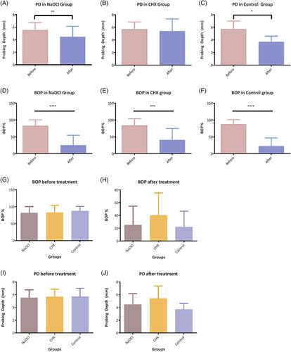
Thirty-five implants were analyzed (CHX: 13; NaOCl: 14; Control:8). NaOCl-treated sites demonstrated reosseointegration with direct bone-to-implant-contact on the previously contaminated surfaces (42% mean reosseointegration), which was significantly higher than Controls (p < 0.05). Correspondingly, clinical improvement was noted with a significant reduction in probing depth from 5.50 ± 1.24 mm at baseline to 4.46 ± 1.70 mm at 6-months postsurgery (p = 0.006). CHX-treated sites demonstrated a nonsignificant reosseointegration of 26% (p > 0.05); however, in the majority of cases, the new bone growth was at a distance from the implant surface without contact. Probing depths did not improve in the CHX group. The use of membrane did not influence reosseointegration or probing depths (all p > 0.05).
Conclusion
Titanium implants with peri-implantitis have the capacity to reosseointegrate following regenerative surgery. However, treatment response is contingent upon the chemotherapeutic agent selection. Additional chemical treatment with 1.5% NaOCl lead to the most favorable results in terms of changes in defect depth and percentage of reosseointegration as compared to CHX, which may hinder reosseointegration.