{"title":"Ultrasonographic images and correspondence with real color sectioned images of the upper limb.","authors":"Seul Ki Kim, Mi-Sun Hur, Jin Seo Park","doi":"10.1007/s00276-024-03410-0","DOIUrl":null,"url":null,"abstract":"<p><strong>Purpose: </strong>For basic training in ultrasonography (US), medical students and residents must learn cross-sectional anatomy. However, the present educational material is not sufficient to learn the sectional anatomy for US. This study aimed to provide a criterion for reading ambiguous structures on US images of upper limb through the sectioned images of Visible Korean.</p><p><strong>Methods: </strong>US images of the right arm of a volunteer were scanned (28 planes). For comparison with US images, the sectioned images of the right upper limb (24 bits color, 0.5 mm intervals, 0.06 mm × 0.06 mm sized pixel) were used. After the volume model was constructed from the sectioned images using MRIcroGL, new sectioned images of 28 planes corresponding to the US images of 28 planes were created by adjusting the slope of the volume model. In all images, the anatomical terms of 59 structures from the shoulder to the fingers were annotated.</p><p><strong>Results: </strong>In the atlas, which consists of 28 sets of US images and sectioned images of various slope planes, 59 structures of the shoulder, arm, elbow, wrist, palm, and fingers were observed in detail.</p><p><strong>Conclusion: </strong>We were able to interpret the ambiguous structures on the US images using the sectioned images with high resolution and actual color. Therefore, to learn the cross-sectional anatomy for US, the sectioned images from the Visible Korean project were deemed to be the suitable data because they contained all human gross anatomical information.</p>","PeriodicalId":49461,"journal":{"name":"Surgical and Radiologic Anatomy","volume":" ","pages":"1469-1479"},"PeriodicalIF":1.4000,"publicationDate":"2024-09-01","publicationTypes":"Journal Article","fieldsOfStudy":null,"isOpenAccess":false,"openAccessPdf":"","citationCount":"0","resultStr":null,"platform":"Semanticscholar","paperid":null,"PeriodicalName":"Surgical and Radiologic Anatomy","FirstCategoryId":"3","ListUrlMain":"https://doi.org/10.1007/s00276-024-03410-0","RegionNum":4,"RegionCategory":"医学","ArticlePicture":[],"TitleCN":null,"AbstractTextCN":null,"PMCID":null,"EPubDate":"2024/6/14 0:00:00","PubModel":"Epub","JCR":"Q2","JCRName":"Medicine","Score":null,"Total":0}
引用次数: 0
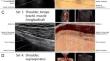
Abstract
Purpose: For basic training in ultrasonography (US), medical students and residents must learn cross-sectional anatomy. However, the present educational material is not sufficient to learn the sectional anatomy for US. This study aimed to provide a criterion for reading ambiguous structures on US images of upper limb through the sectioned images of Visible Korean.
Methods: US images of the right arm of a volunteer were scanned (28 planes). For comparison with US images, the sectioned images of the right upper limb (24 bits color, 0.5 mm intervals, 0.06 mm × 0.06 mm sized pixel) were used. After the volume model was constructed from the sectioned images using MRIcroGL, new sectioned images of 28 planes corresponding to the US images of 28 planes were created by adjusting the slope of the volume model. In all images, the anatomical terms of 59 structures from the shoulder to the fingers were annotated.
Results: In the atlas, which consists of 28 sets of US images and sectioned images of various slope planes, 59 structures of the shoulder, arm, elbow, wrist, palm, and fingers were observed in detail.
Conclusion: We were able to interpret the ambiguous structures on the US images using the sectioned images with high resolution and actual color. Therefore, to learn the cross-sectional anatomy for US, the sectioned images from the Visible Korean project were deemed to be the suitable data because they contained all human gross anatomical information.
目的:医科学生和住院医师在接受超声造影术(US)基础培训时,必须学习横断面解剖。然而,目前的教材不足以学习 US 的断面解剖。本研究旨在通过韩国可见光切面图像,为阅读上肢 US 图像上的模糊结构提供一个标准:方法:扫描一名志愿者右臂的 US 图像(28 个平面)。为了与 US 图像进行比较,使用了右上肢的切面图像(24 位彩色、0.5 毫米间隔、0.06 毫米 × 0.06 毫米大小的像素)。使用 MRIcroGL 根据切片图像构建容积模型后,通过调整容积模型的斜率,创建了与 US 图像 28 个平面相对应的 28 个平面的新切片图像。在所有图像中,标注了从肩部到手指的 59 个结构的解剖术语:结果:在由 28 组 US 图像和不同斜面的切片图像组成的图集中,详细观察了肩、臂、肘、腕、掌和手指的 59 个结构:结论:我们能够利用高分辨率和真实色彩的切面图像解读 US 图像上的模糊结构。因此,要学习 US 截面解剖,"看得见的韩国 "项目的截面图像被认为是合适的数据,因为它们包含了所有的人体大体解剖信息。
期刊介绍:
Anatomy is a morphological science which cannot fail to interest the clinician. The practical application of anatomical research to clinical problems necessitates special adaptation and selectivity in choosing from numerous international works. Although there is a tendency to believe that meaningful advances in anatomy are unlikely, constant revision is necessary. Surgical and Radiologic Anatomy, the first international journal of Clinical anatomy has been created in this spirit.
Its goal is to serve clinicians, regardless of speciality-physicians, surgeons, radiologists or other specialists-as an indispensable aid with which they can improve their knowledge of anatomy. Each issue includes: Original papers, review articles, articles on the anatomical bases of medical, surgical and radiological techniques, articles of normal radiologic anatomy, brief reviews of anatomical publications of clinical interest.
Particular attention is given to high quality illustrations, which are indispensable for a better understanding of anatomical problems.
Surgical and Radiologic Anatomy is a journal written by anatomists for clinicians with a special interest in anatomy.

 求助内容:
求助内容: 应助结果提醒方式:


