Developmental morpho-analysis of the caecum in Japanese quail embryos (Coturnix coturnix japonica)

IF 2 3区 工程技术 Q2 ANATOMY & MORPHOLOGY
Fatma Abdelhakeem
{"title":"Developmental morpho-analysis of the caecum in Japanese quail embryos (Coturnix coturnix japonica)","authors":"Fatma Abdelhakeem","doi":"10.1002/jemt.24632","DOIUrl":null,"url":null,"abstract":"<div>\n \n \n <section>\n \n <p>In the current study, we are focusing on the microanatomical structure of quail caecum during the prehatching time to try to understand the function and the role of each cell-built quail caecum reaching how caecum plays an essential role in immunity and absorption. The morpho-developmental features of the quail caecum were described in detail daily from the third incubation day (ID) till hatching time, investigating the gross morphology, microscopic, and ultrastructure using light and scanning electron microscope. The embryonic caecum appeared grossly as two lateral outpocketings with blinded ends, emerging laterally at the junction between the small and large intestine (the ileocaecal junction). The primordia of two caeca, represented by two lateral swellings from the hindgut on the fourth ID, continued growing till the day of hatching, where the caecal wall consisted of three apparent layers: mucosa, musculosa, and serosa. At the time of hatching, the quail caecum was still not fully mature and will continue growing posthatching. The findings in this study can be applied in further studies intended to understand the physiological mechanisms of the caecum during prehatching and posthatching periods.</p>\n </section>\n \n <section>\n \n <h3> Research Highlights</h3>\n \n <div>\n <ul>\n \n <li>Caecum is one of the hindgut derivatives that started as two lateral swellings.</li>\n \n <li>The caecal wall consisted of three layers; mucosa, musculosa, and serosa.</li>\n \n <li>The caecum plays an essential role in immunity maintenance.</li>\n \n <li>Caecum continues to grow posthatching as it is not fully mature at hatching time.</li>\n </ul>\n </div>\n </section>\n </div>","PeriodicalId":18684,"journal":{"name":"Microscopy Research and Technique","volume":null,"pages":null},"PeriodicalIF":2.0000,"publicationDate":"2024-06-12","publicationTypes":"Journal Article","fieldsOfStudy":null,"isOpenAccess":false,"openAccessPdf":"","citationCount":"0","resultStr":null,"platform":"Semanticscholar","paperid":null,"PeriodicalName":"Microscopy Research and Technique","FirstCategoryId":"5","ListUrlMain":"https://onlinelibrary.wiley.com/doi/10.1002/jemt.24632","RegionNum":3,"RegionCategory":"工程技术","ArticlePicture":[],"TitleCN":null,"AbstractTextCN":null,"PMCID":null,"EPubDate":"","PubModel":"","JCR":"Q2","JCRName":"ANATOMY & MORPHOLOGY","Score":null,"Total":0}
引用次数: 0

Abstract

In the current study, we are focusing on the microanatomical structure of quail caecum during the prehatching time to try to understand the function and the role of each cell-built quail caecum reaching how caecum plays an essential role in immunity and absorption. The morpho-developmental features of the quail caecum were described in detail daily from the third incubation day (ID) till hatching time, investigating the gross morphology, microscopic, and ultrastructure using light and scanning electron microscope. The embryonic caecum appeared grossly as two lateral outpocketings with blinded ends, emerging laterally at the junction between the small and large intestine (the ileocaecal junction). The primordia of two caeca, represented by two lateral swellings from the hindgut on the fourth ID, continued growing till the day of hatching, where the caecal wall consisted of three apparent layers: mucosa, musculosa, and serosa. At the time of hatching, the quail caecum was still not fully mature and will continue growing posthatching. The findings in this study can be applied in further studies intended to understand the physiological mechanisms of the caecum during prehatching and posthatching periods.

Research Highlights

  • Caecum is one of the hindgut derivatives that started as two lateral swellings.
  • The caecal wall consisted of three layers; mucosa, musculosa, and serosa.
  • The caecum plays an essential role in immunity maintenance.
  • Caecum continues to grow posthatching as it is not fully mature at hatching time.
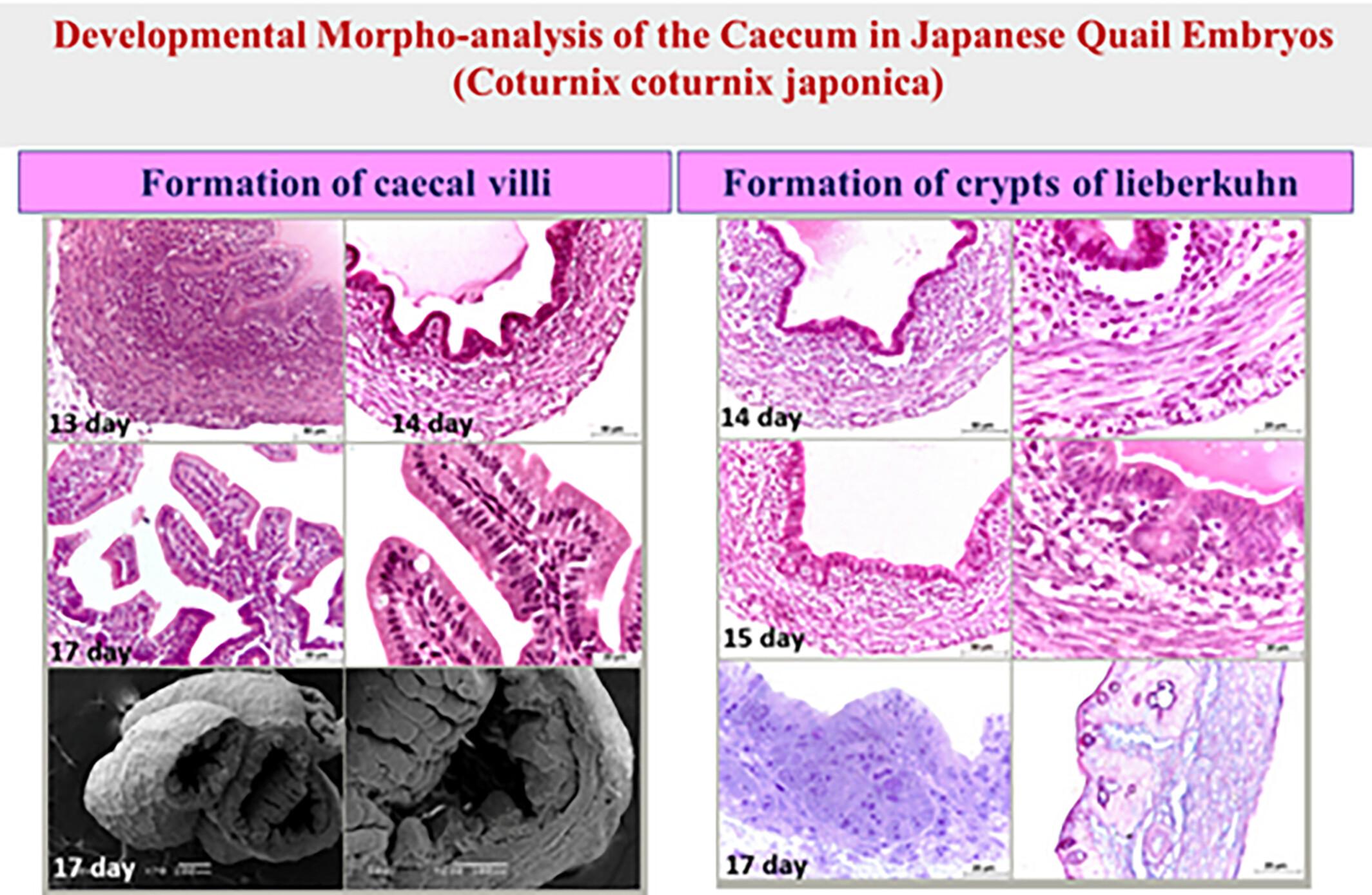
Abstract Image

日本鹌鹑胚胎盲肠的发育形态分析
在本研究中,我们关注鹌鹑盲肠在孵化前的微观解剖结构,试图了解每个细胞构建的鹌鹑盲肠的功能和作用,以及盲肠如何在免疫和吸收中发挥重要作用。从孵化第三天(ID)到孵化前,每天都对鹌鹑盲肠的形态发育特征进行了详细描述,并使用光学显微镜和扫描电子显微镜对其大体形态、显微镜和超微结构进行了研究。胚胎盲肠的大体形态为两侧外翻,末端有盲端,在小肠和大肠的交界处(回盲肠交界处)横向出现。两个盲肠的原基由第四个ID上来自后肠的两个侧向肿物代表,一直生长到孵化当天,盲肠壁由三个明显的层组成:粘膜层、肌肉层和浆膜层。孵化时,鹌鹑的盲肠仍未完全成熟,孵化后将继续生长。本研究的结果可用于进一步研究,以了解盲肠在孵化前和孵化后的生理机制。研究亮点:盲肠是后肠衍生物之一,最初是两个侧向肿胀。盲肠壁由粘膜、肌层和浆膜三层组成。盲肠在维持免疫力方面发挥着重要作用。盲肠在孵化后继续生长,因为它在孵化时尚未完全成熟。
本文章由计算机程序翻译,如有差异,请以英文原文为准。
求助全文
约1分钟内获得全文 求助全文
来源期刊
Microscopy Research and Technique
Microscopy Research and Technique 医学-解剖学与形态学
CiteScore
5.30
自引率
20.00%
发文量
233
审稿时长
4.7 months
期刊介绍: Microscopy Research and Technique (MRT) publishes articles on all aspects of advanced microscopy original architecture and methodologies with applications in the biological, clinical, chemical, and materials sciences. Original basic and applied research as well as technical papers dealing with the various subsets of microscopy are encouraged. MRT is the right form for those developing new microscopy methods or using the microscope to answer key questions in basic and applied research.
×
引用
GB/T 7714-2015
复制
MLA
复制
APA
复制
导出至
BibTeX EndNote RefMan NoteFirst NoteExpress
×
提示
您的信息不完整,为了账户安全,请先补充。
现在去补充
×
提示
您因"违规操作"
具体请查看互助需知
我知道了
×
提示
确定
请完成安全验证×
copy
已复制链接
快去分享给好友吧!
我知道了
右上角分享
点击右上角分享
0
联系我们:info@booksci.cn Book学术提供免费学术资源搜索服务,方便国内外学者检索中英文文献。致力于提供最便捷和优质的服务体验。 Copyright © 2023 布克学术 All rights reserved.
京ICP备2023020795号-1
ghs 京公网安备 11010802042870号
Book学术文献互助
Book学术文献互助群
群 号:481959085
Book学术官方微信