{"title":"Review of the role of imaging in the diagnosis of priapism.","authors":"Conrad von Stempel, Miles Walkden, Alex Kirkham","doi":"10.1038/s41443-024-00928-0","DOIUrl":null,"url":null,"abstract":"<p><p>Imaging has a specific role in the diagnosis and management of priapism. The primary imaging modality is ultrasound with colour Doppler (CDUS) which can accurately assess the hemodynamics of the cavernosal arteries. This is particularly useful in equivocal cases and can help differentiate ischemic from non-ischemic priapism as well as confirm the presence and location of arterio-venous fistulae post penile trauma. Furthermore, CDUS is invaluable in the post treatment follow up of non-ischemic priapism. Contrast enhanced magnetic resonance imaging (MRI) can demonstrate the extent of cavernosal necrosis in ischemic priapism and in conjunction with computer tomography (CT) has an important role in excluding underlying malignancy. MRI and CT angiography are used to evaluate pudendal arterial anatomy, which can be extremely variable and aids in the management of non-ischemic priapism. In selected cases of non-ischemic priapism, catheter angiography and transcatheter embolization of arteriovenous fistulae is an effective treatment. This review will examine the specific roles of different imaging modalities in the subtypes of priapism as well as highlight some of the pitfalls encountered in imaging.</p>","PeriodicalId":14068,"journal":{"name":"International Journal of Impotence Research","volume":null,"pages":null},"PeriodicalIF":2.8000,"publicationDate":"2024-06-11","publicationTypes":"Journal Article","fieldsOfStudy":null,"isOpenAccess":false,"openAccessPdf":"","citationCount":"0","resultStr":null,"platform":"Semanticscholar","paperid":null,"PeriodicalName":"International Journal of Impotence Research","FirstCategoryId":"3","ListUrlMain":"https://doi.org/10.1038/s41443-024-00928-0","RegionNum":3,"RegionCategory":"医学","ArticlePicture":[],"TitleCN":null,"AbstractTextCN":null,"PMCID":null,"EPubDate":"","PubModel":"","JCR":"Q2","JCRName":"UROLOGY & NEPHROLOGY","Score":null,"Total":0}
引用次数: 0
Abstract
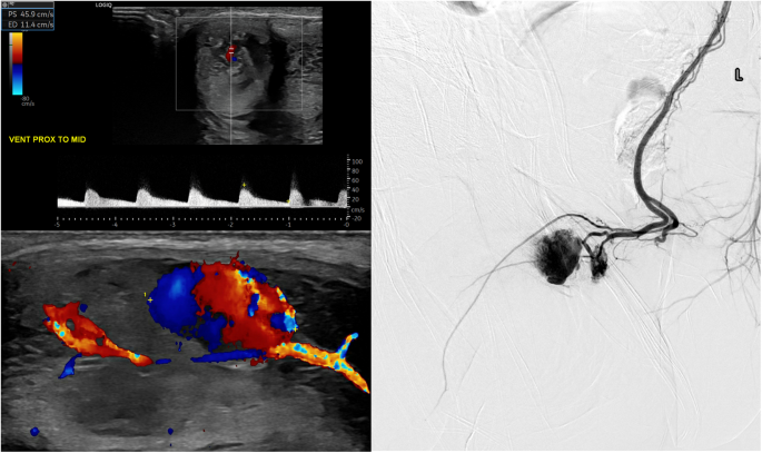
Imaging has a specific role in the diagnosis and management of priapism. The primary imaging modality is ultrasound with colour Doppler (CDUS) which can accurately assess the hemodynamics of the cavernosal arteries. This is particularly useful in equivocal cases and can help differentiate ischemic from non-ischemic priapism as well as confirm the presence and location of arterio-venous fistulae post penile trauma. Furthermore, CDUS is invaluable in the post treatment follow up of non-ischemic priapism. Contrast enhanced magnetic resonance imaging (MRI) can demonstrate the extent of cavernosal necrosis in ischemic priapism and in conjunction with computer tomography (CT) has an important role in excluding underlying malignancy. MRI and CT angiography are used to evaluate pudendal arterial anatomy, which can be extremely variable and aids in the management of non-ischemic priapism. In selected cases of non-ischemic priapism, catheter angiography and transcatheter embolization of arteriovenous fistulae is an effective treatment. This review will examine the specific roles of different imaging modalities in the subtypes of priapism as well as highlight some of the pitfalls encountered in imaging.
期刊介绍:
International Journal of Impotence Research: The Journal of Sexual Medicine addresses sexual medicine for both genders as an interdisciplinary field. This includes basic science researchers, urologists, endocrinologists, cardiologists, family practitioners, gynecologists, internists, neurologists, psychiatrists, psychologists, radiologists and other health care clinicians.
 求助内容:
求助内容: 应助结果提醒方式:


