First report of the emerging rosette agent (Sphaerothecum destruens) in a captive held native north American cyprinid, the warpaint shiner (Luxilus coccogenis, Cope)
Bridgette K. Gunn, John H. Leary, Vivian M. Lee, Ashley J. Kirby, Gregory Scott, Alvin C. Camus
{"title":"First report of the emerging rosette agent (Sphaerothecum destruens) in a captive held native north American cyprinid, the warpaint shiner (Luxilus coccogenis, Cope)","authors":"Bridgette K. Gunn, John H. Leary, Vivian M. Lee, Ashley J. Kirby, Gregory Scott, Alvin C. Camus","doi":"10.1111/jfd.13980","DOIUrl":null,"url":null,"abstract":"<p><i>Sphaerothecum destruens</i>, the rosette agent, is a unicellular, obligate intracellular, mesomycetozoan pathogen at the phylogenetic intersection between animals and fungi (Combe et al., <span>2022</span>; Paley et al., <span>2012</span>). First associated with diseased North American (NA) salmonids, the parasite has increasingly been documented in European cyprinids where spread is attributed to introduction of the invasive East Asian topmouth gudgeon (<i>Pseudorasbora parva</i>) (Andreou et al., <span>2011</span>; Arkush et al., <span>1998</span>; Combe et al., <span>2022</span>; Combe & Gozlan, <span>2018</span>; Spikmans et al., <span>2019</span>). Impacts of the parasite can be high, with mortalities exceeding 80% in diseased net pen reared chinook salmon (<i>Onchorhynchus tshawytscha</i>) (Harrell et al., <span>1986</span>). However, infected chinook have lived 3–5 years and spawned successfully (Arkush et al., <span>1998</span>). Losses among susceptible wild fish species are more difficult to document and the disease appears more insidious, resulting in low-level mortalities and population declines over time, including the disappearance of sunbleak (<i>Leucaspius delineates</i>) from much of Europe (Gozlan & Combe, <span>2023</span>).</p><p>Formerly a D.R.I.P. clade member (Ragan et al., <span>1996</span>), class Ichthyosporea, organisms were grouped according to shared morphological features and 18S rRNA molecular analyses (Gozlan & Combe, <span>2023</span>). Members included <i>Dermocystidium</i> spp., the rosette agent, <i>Ichthyophonus hoferi</i>, and <i>Psorospermium haeckeli</i> (Ragan et al., <span>1996</span>). Subsequent phylogenetic investigation reclassified the group under the class Mesomycetozoa (Herr et al., <span>1999</span>; Mendoza et al., <span>2001</span>, <span>2002</span>), placing <i>S. destruens</i>, <i>Dermocystidium</i> spp. and <i>Rhinosporidium seeberi</i> in the order Dermocystida (Cavalier-Smith, <span>1998</span>; Gozlan & Combe, <span>2023</span>). Today, <i>S. destruens</i> is within the super-group Opisthokonta with eukaryotic fungi, choanoflagellates and animals (Gozlan & Combe, <span>2023</span>).</p><p>The <i>S. destruens</i> lifecycle is poorly understood but includes distinctive 2–4 μm (undivided) and 4–6 μm (dividing) spores, and 2 μm uniflagellate zoospores (Arkush et al., <span>1998</span>; Gozlan & Combe, <span>2023</span>). Infection presumptively occurs through ingestion or adherence of motile zoospores to gills or skin, followed by asexual replication intracellularly in host tissues. Disease can be disseminated or nodular, corresponding to necrotizing lesions with numerous eosinophilic spores and little inflammation or with spores limited to granulomas primarily within visceral organs, respectively (Arkush et al., <span>1998</span>). Cell rupture ultimately releases spores via bodily fluids. Zoosporulation, completed in freshwater, exhibits broad temperature tolerance (Andreou et al., <span>2009</span>, <span>2011</span>; Arkush et al., <span>2003</span>; Gozlan & Combe, <span>2023</span>).</p><p>Biodiversity among freshwater fishes is threatened by habitat availability and anthropogenic activity. Emerging infectious agents, such as <i>S. destruens</i>, can exacerbate these threats and devastate naïve indigenous populations (Daszak et al., <span>2000</span>; Gozlan & Combe, <span>2023</span>). Complicating epidemiological factors is the potential for introductions of silent carrier species that serve as environmental reservoirs and drivers of infection. Spread of <i>S. destruens</i> among European cyprinids following introduction of the topmouth gudgeon, exemplifies this (Andreou et al., <span>2011</span>; Combe et al., <span>2022</span>; Combe & Gozlan, <span>2018</span>; Spikmans et al., <span>2019</span>). This report describes histopathological, electron microscopic and in situ hybridization findings in the first documented case of <i>S. destruens</i> infection in a native NA cyprinid, the warpaint shiner (<i>Luxilus coccogenis</i>, Cope). While the source of infection in this case could not be determined, identification of the parasite raises concerns for the health of native cyprinid populations in NA.</p><p>Formalin-fixed tissues from an approximately 10 cm warpaint shiner, age and sex unknown, were received by the Aquatic Pathology Service, University of Georgia, Athens, Georgia in May 2023. Originally collected in Cooper Creek, Union County, GA, USA (34.761805, −84.090040), the shiner was displayed in a public aquarium with other native cyprinids [longnose dace (<i>Rhinichthys cataractae</i>), mirror shiner (<i>Notropis spectrunculus</i>), rainbow shiner (<i>N. chrosomus</i>), Tennessee shiner (<i>N. leuciodus</i>)] and non-cyprinid [gilt darter (<i>Percina evides</i>), greenside darter (<i>Etheostoma blennioides</i>), redline darter (<i>Etheostoma rufilineatum</i>), sculpin (<i>Cottus</i> sp.)] species at a constant water temperature of 20°C. Found acutely moribund, the fish was euthanized in 500 mg/L buffered tricaine methanesulfonate and necropsied by aquarium personnel. Tissues were processed routinely for histological evaluation and stained with haematoxylin and eosin (H&E), modified Brown and Hopps (BH), periodic acid-Schiff (PAS), Gomori's methenamine silver (GMS), and Ziehl-Neelsen (ZN) stains. For transmission electron microscopy, 1–2 mm cubes of formalin-fixed liver were transferred to modified Karnovsky's solution, processed using standard methods, and observed with a JEOL JEM101 transmission electron microscope.</p><p>Identification of <i>S. destruens</i> in histological sections utilized RNAscope® in situ hybridization (ISH) (Advanced Cell Diagnostics Inc., Hayward, CA) (Wang et al., <span>2012</span>). Alignment of multiple <i>S. destruens</i> 18S rRNA sequences in the NCBI nucleotide database with outgroup species (<i>Dermocystidium</i> spp., <i>Ichthyophonus hoferi, Rhinosporidium seeberi</i>) identified a highly conserved region useful for RNAscope probe design. A 74 bp probe suitable for ISH (CGCCGCGAGGTGTTTGCCCCGACGAGGGTGATCCTTCCTCTCGAAATTGGCGTGTGCGCTTAATTGAGTGTGCG) was synthesized and tested for specificity to <i>S. destruens</i>.</p><p>The hybridization assay followed the RNAscope® 2.5 HD Detection Kit (RED) protocol. Unstained histological sections on charged slides were deparaffinized, dried, treated with hydrogen peroxide, rinsed with distilled water and immersed in target retrieval solution (99°C, 15 min). Stock <i>S. destruens</i> probe or RNAscope® Negative Control Probe (DapB) was added and incubated (40°C, 2 h). Detection steps were as per protocol. Slides were counterstained with haematoxylin followed by nuclear bluing (0.02% ammonium hydroxide) and coverslipping. Potential probe cross-reactivity with non-target mesomycetozoans and host tissue was assessed using histological sections from a redspot darter (<i>Etheostoma artesiae</i>) and brook trout (<i>Salvelinus fontinalis</i>) containing lesions histologically consistent with a <i>Dermocystidium</i> sp. and <i>I. hoferi</i>, respectively. Bighead carp (<i>Hypophthalmichthys nobilis</i>) tissues confirmed positive for <i>S. destruens</i> by PCR served as a positive control (González-Hernández et al., <span>2010</span>).</p><p>Necropsy findings included with the clinical history indicated a few gill monogeneans and a subjectively enlarged liver and posterior kidney. The cranial liver, sharply demarcated from the normal posterior half, was friable, mottled pink to tan, and contained a 0.5 cm dark red focus. With routine H&E staining of histological sections, multiple foci of hepatocellular necrosis were associated with intracellular and occasional extracellular, round, 2–6 μm, magenta spores occurring individually or in clusters (Figure 1a). Additional spores were observed rarely within macrophage aggregates and discrete granulomas (Figure 1b), as well as within biliary epithelial and endothelial cells. Spores were PAS and GMS positive (Figure 1c,d), acid-fast (ZN) negative and Gram (BH) variable. A few necrotic foci and granulomas were scattered in renal haematopoietic areas. Rare spores were present in renal tubular lumens and epithelial cells, the endocardium, peripancreatic adipose, intestinal mucosa and macrophages within branchial central venous sinuses. Groups of unidentified round to ovoid, 1–2 μm organisms with intensely basophilic nuclei and thin rims of clear cytoplasm were also infrequently observed in hepatocyte cytoplasm on the periphery of necrotic foci (Figure 1d). Additional findings included ulcerative dermatitis and branchial saprolegniasis.</p><p>Electron microscopy revealed only nondividing spores. A trilaminar plasma membrane (Figure 2a) surrounded granular cytoplasm with large numbers of free ribosomes, scattered rough endoplasmic reticulum, vesicular bodies, concentric bodies, electron-dense and electron-lucent bodies. Spores were usually within large membrane-bound structures of presumed host cell origin mixed with abundant debris and degenerate spores (Figure 2b).</p><p>The ISH probe strongly labelled spores in the case shiner and carp positive control (Figure 3a,b). There was no cross-reactivity with any fish tissues, <i>Dermocystidium</i> sp. or <i>I. hoferi</i>. No staining occurred in the negative control (Figure 3c,d).</p><p>Microscopic findings in this warpaint shiner, including clusters of small, round, magenta stained spores within areas of necrosis and granulomas in H&E stained tissue sections, are consistent with morphological descriptions of, and lesions induced by, <i>S. destruens</i>, the ‘rosette agent’. In addition, spores stained positively with PAS (magenta) and GMS (black) histochemical stains demonstrating the presence of polysaccharides in their cell walls, a reflection of the relationship of mesomycetozoans to fungi (Figure 1a–d) (Andreou et al., <span>2011</span>; Arkush et al., <span>1998</span>; Gozlan & Combe, <span>2023</span>). Confirmatory PCR was not attempted as only formalin-fixed paraffin-embedded tissues were available. However, RNAScope® ISH positively labelled intralesional <i>S. destruens</i> spores. The unidentified intracellular organism did not label with the ISH probe (Figure 1e).</p><p>Originally described from salmonids in the northwestern United States (US) (Elston et al., <span>1986</span>; Harrell et al., <span>1986</span>), <i>S. destruens</i> has caused disease in increasing numbers of salmonid and non-salmonid, including cyprinid, species in Europe since 2005 (Gozlan & Combe, <span>2023</span>). This is the first report of <i>S. destruens</i> infection in a native NA cyprinid, and in the southeastern US. However, questions remain concerning the source and timing of infection in this case, as well as its potential significance to US cyprinids. Although the shiner originated in a North Georgia waterway, it was also displayed in a mixed species aquarium exhibit for over 20 months at 20°C, confounding whether infection was acquired in the natural or aquarium environment. While the temperature was above the 15°C optimum for zoosporulation, it was still conducive to high zoospore concentrations in tank water and potentially to disease transmission (Andreou et al., <span>2009</span>). While infected salmonids can survive 3–5 years, suggesting the shiner could have been infected prior to collection, another species of native fish could have acted as a carrier of the parasite in the aquarium population (Arkush et al., <span>1998</span>). Despite this, no evidence of infection has been previously detected among 30 mortalities examined histologically from the mixed species exhibit since 2007. However, while six additional mortalities occurred among warpaint shiners following the case submission, due to autolysis only one was suitable for histopathological examination suggesting evidence of wider infection could have been missed.</p><p>In their native Tennessee River drainage range and introduced southeastern sites, warpaint shiners could potentially encounter sources of infection in rainbow (<i>Oncorhynchus mykiss</i>) and brown (<i>Salmo trutta</i>) trout (Gozlan & Combe, <span>2023</span>; Nico & Fuller, <span>2019</span>). Alternatively, an unknown natural or introduced infective reservoir could exist. Translocation of Asian carp into Europe inadvertently introduced the topmouth gudgeon carrier, resulting in severe declines of native cyprinid populations (Combe et al., <span>2022</span>; Combe & Gozlan, <span>2018</span>; Fusaro et al., <span>2022</span>). Should additional cases arise, strain determination through sequencing of the ITS-1 genetic marker could help localize its origin. American strains are genetically related, while European and Asian strains are more diverse (Combe & Gozlan, <span>2018</span>).</p><p>The significance of this isolated case is unclear, although identification of the rosette agent in a native NA cyprinid is concerning, meriting further investigation and increased awareness within the fish health community. While mortalities vary, depending on host physiological condition and environmental stressors, they can approach 100% (Arkush et al., <span>1998</span>; Gozlan & Combe, <span>2023</span>). Despite this, population declines in natural environments can go unnoticed, particularly with small fish species. In Europe, <i>S. destruens</i> existed unidentified for over 45 years, despite diminishing cyprinid numbers and biodiversity (Combe & Gozlan, <span>2018</span>). As a result, epidemiological studies combining molecular, histopathological and ecological monitoring are warranted to better characterize <i>S. destruens</i> distribution and host range in NA freshwater fishes.</p><p>Bridgette Gunn: Conceptualization; investigation; writing – original draft; writing – review and editing. John Leary: investigation; supervision; validation. Vivian Lee: Investigation; resources; writing – review and editing. Ashley Kirby: Investigation; resources; writing – review and editing. Gregory Scott: Investigation; resources; writing – review and editing. Alvin Camus: Conceptualization; investigation; resources; supervision; validation; writing – original draft; writing – review and editing.</p><p>None.</p><p>The authors declare no conflicts of interests related to the performance or publication of the work described herein.</p>","PeriodicalId":15849,"journal":{"name":"Journal of fish diseases","volume":"47 9","pages":""},"PeriodicalIF":2.2000,"publicationDate":"2024-06-10","publicationTypes":"Journal Article","fieldsOfStudy":null,"isOpenAccess":false,"openAccessPdf":"https://onlinelibrary.wiley.com/doi/epdf/10.1111/jfd.13980","citationCount":"0","resultStr":null,"platform":"Semanticscholar","paperid":null,"PeriodicalName":"Journal of fish diseases","FirstCategoryId":"97","ListUrlMain":"https://onlinelibrary.wiley.com/doi/10.1111/jfd.13980","RegionNum":3,"RegionCategory":"农林科学","ArticlePicture":[],"TitleCN":null,"AbstractTextCN":null,"PMCID":null,"EPubDate":"","PubModel":"","JCR":"Q2","JCRName":"FISHERIES","Score":null,"Total":0}
引用次数: 0
Abstract
Sphaerothecum destruens, the rosette agent, is a unicellular, obligate intracellular, mesomycetozoan pathogen at the phylogenetic intersection between animals and fungi (Combe et al., 2022; Paley et al., 2012). First associated with diseased North American (NA) salmonids, the parasite has increasingly been documented in European cyprinids where spread is attributed to introduction of the invasive East Asian topmouth gudgeon (Pseudorasbora parva) (Andreou et al., 2011; Arkush et al., 1998; Combe et al., 2022; Combe & Gozlan, 2018; Spikmans et al., 2019). Impacts of the parasite can be high, with mortalities exceeding 80% in diseased net pen reared chinook salmon (Onchorhynchus tshawytscha) (Harrell et al., 1986). However, infected chinook have lived 3–5 years and spawned successfully (Arkush et al., 1998). Losses among susceptible wild fish species are more difficult to document and the disease appears more insidious, resulting in low-level mortalities and population declines over time, including the disappearance of sunbleak (Leucaspius delineates) from much of Europe (Gozlan & Combe, 2023).
Formerly a D.R.I.P. clade member (Ragan et al., 1996), class Ichthyosporea, organisms were grouped according to shared morphological features and 18S rRNA molecular analyses (Gozlan & Combe, 2023). Members included Dermocystidium spp., the rosette agent, Ichthyophonus hoferi, and Psorospermium haeckeli (Ragan et al., 1996). Subsequent phylogenetic investigation reclassified the group under the class Mesomycetozoa (Herr et al., 1999; Mendoza et al., 2001, 2002), placing S. destruens, Dermocystidium spp. and Rhinosporidium seeberi in the order Dermocystida (Cavalier-Smith, 1998; Gozlan & Combe, 2023). Today, S. destruens is within the super-group Opisthokonta with eukaryotic fungi, choanoflagellates and animals (Gozlan & Combe, 2023).
The S. destruens lifecycle is poorly understood but includes distinctive 2–4 μm (undivided) and 4–6 μm (dividing) spores, and 2 μm uniflagellate zoospores (Arkush et al., 1998; Gozlan & Combe, 2023). Infection presumptively occurs through ingestion or adherence of motile zoospores to gills or skin, followed by asexual replication intracellularly in host tissues. Disease can be disseminated or nodular, corresponding to necrotizing lesions with numerous eosinophilic spores and little inflammation or with spores limited to granulomas primarily within visceral organs, respectively (Arkush et al., 1998). Cell rupture ultimately releases spores via bodily fluids. Zoosporulation, completed in freshwater, exhibits broad temperature tolerance (Andreou et al., 2009, 2011; Arkush et al., 2003; Gozlan & Combe, 2023).
Biodiversity among freshwater fishes is threatened by habitat availability and anthropogenic activity. Emerging infectious agents, such as S. destruens, can exacerbate these threats and devastate naïve indigenous populations (Daszak et al., 2000; Gozlan & Combe, 2023). Complicating epidemiological factors is the potential for introductions of silent carrier species that serve as environmental reservoirs and drivers of infection. Spread of S. destruens among European cyprinids following introduction of the topmouth gudgeon, exemplifies this (Andreou et al., 2011; Combe et al., 2022; Combe & Gozlan, 2018; Spikmans et al., 2019). This report describes histopathological, electron microscopic and in situ hybridization findings in the first documented case of S. destruens infection in a native NA cyprinid, the warpaint shiner (Luxilus coccogenis, Cope). While the source of infection in this case could not be determined, identification of the parasite raises concerns for the health of native cyprinid populations in NA.
Formalin-fixed tissues from an approximately 10 cm warpaint shiner, age and sex unknown, were received by the Aquatic Pathology Service, University of Georgia, Athens, Georgia in May 2023. Originally collected in Cooper Creek, Union County, GA, USA (34.761805, −84.090040), the shiner was displayed in a public aquarium with other native cyprinids [longnose dace (Rhinichthys cataractae), mirror shiner (Notropis spectrunculus), rainbow shiner (N. chrosomus), Tennessee shiner (N. leuciodus)] and non-cyprinid [gilt darter (Percina evides), greenside darter (Etheostoma blennioides), redline darter (Etheostoma rufilineatum), sculpin (Cottus sp.)] species at a constant water temperature of 20°C. Found acutely moribund, the fish was euthanized in 500 mg/L buffered tricaine methanesulfonate and necropsied by aquarium personnel. Tissues were processed routinely for histological evaluation and stained with haematoxylin and eosin (H&E), modified Brown and Hopps (BH), periodic acid-Schiff (PAS), Gomori's methenamine silver (GMS), and Ziehl-Neelsen (ZN) stains. For transmission electron microscopy, 1–2 mm cubes of formalin-fixed liver were transferred to modified Karnovsky's solution, processed using standard methods, and observed with a JEOL JEM101 transmission electron microscope.
Identification of S. destruens in histological sections utilized RNAscope® in situ hybridization (ISH) (Advanced Cell Diagnostics Inc., Hayward, CA) (Wang et al., 2012). Alignment of multiple S. destruens 18S rRNA sequences in the NCBI nucleotide database with outgroup species (Dermocystidium spp., Ichthyophonus hoferi, Rhinosporidium seeberi) identified a highly conserved region useful for RNAscope probe design. A 74 bp probe suitable for ISH (CGCCGCGAGGTGTTTGCCCCGACGAGGGTGATCCTTCCTCTCGAAATTGGCGTGTGCGCTTAATTGAGTGTGCG) was synthesized and tested for specificity to S. destruens.
The hybridization assay followed the RNAscope® 2.5 HD Detection Kit (RED) protocol. Unstained histological sections on charged slides were deparaffinized, dried, treated with hydrogen peroxide, rinsed with distilled water and immersed in target retrieval solution (99°C, 15 min). Stock S. destruens probe or RNAscope® Negative Control Probe (DapB) was added and incubated (40°C, 2 h). Detection steps were as per protocol. Slides were counterstained with haematoxylin followed by nuclear bluing (0.02% ammonium hydroxide) and coverslipping. Potential probe cross-reactivity with non-target mesomycetozoans and host tissue was assessed using histological sections from a redspot darter (Etheostoma artesiae) and brook trout (Salvelinus fontinalis) containing lesions histologically consistent with a Dermocystidium sp. and I. hoferi, respectively. Bighead carp (Hypophthalmichthys nobilis) tissues confirmed positive for S. destruens by PCR served as a positive control (González-Hernández et al., 2010).
Necropsy findings included with the clinical history indicated a few gill monogeneans and a subjectively enlarged liver and posterior kidney. The cranial liver, sharply demarcated from the normal posterior half, was friable, mottled pink to tan, and contained a 0.5 cm dark red focus. With routine H&E staining of histological sections, multiple foci of hepatocellular necrosis were associated with intracellular and occasional extracellular, round, 2–6 μm, magenta spores occurring individually or in clusters (Figure 1a). Additional spores were observed rarely within macrophage aggregates and discrete granulomas (Figure 1b), as well as within biliary epithelial and endothelial cells. Spores were PAS and GMS positive (Figure 1c,d), acid-fast (ZN) negative and Gram (BH) variable. A few necrotic foci and granulomas were scattered in renal haematopoietic areas. Rare spores were present in renal tubular lumens and epithelial cells, the endocardium, peripancreatic adipose, intestinal mucosa and macrophages within branchial central venous sinuses. Groups of unidentified round to ovoid, 1–2 μm organisms with intensely basophilic nuclei and thin rims of clear cytoplasm were also infrequently observed in hepatocyte cytoplasm on the periphery of necrotic foci (Figure 1d). Additional findings included ulcerative dermatitis and branchial saprolegniasis.
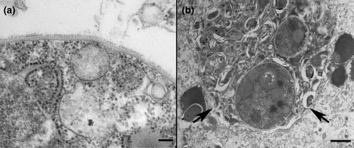
Electron microscopy revealed only nondividing spores. A trilaminar plasma membrane (Figure 2a) surrounded granular cytoplasm with large numbers of free ribosomes, scattered rough endoplasmic reticulum, vesicular bodies, concentric bodies, electron-dense and electron-lucent bodies. Spores were usually within large membrane-bound structures of presumed host cell origin mixed with abundant debris and degenerate spores (Figure 2b).
The ISH probe strongly labelled spores in the case shiner and carp positive control (Figure 3a,b). There was no cross-reactivity with any fish tissues, Dermocystidium sp. or I. hoferi. No staining occurred in the negative control (Figure 3c,d).
Microscopic findings in this warpaint shiner, including clusters of small, round, magenta stained spores within areas of necrosis and granulomas in H&E stained tissue sections, are consistent with morphological descriptions of, and lesions induced by, S. destruens, the ‘rosette agent’. In addition, spores stained positively with PAS (magenta) and GMS (black) histochemical stains demonstrating the presence of polysaccharides in their cell walls, a reflection of the relationship of mesomycetozoans to fungi (Figure 1a–d) (Andreou et al., 2011; Arkush et al., 1998; Gozlan & Combe, 2023). Confirmatory PCR was not attempted as only formalin-fixed paraffin-embedded tissues were available. However, RNAScope® ISH positively labelled intralesional S. destruens spores. The unidentified intracellular organism did not label with the ISH probe (Figure 1e).
Originally described from salmonids in the northwestern United States (US) (Elston et al., 1986; Harrell et al., 1986), S. destruens has caused disease in increasing numbers of salmonid and non-salmonid, including cyprinid, species in Europe since 2005 (Gozlan & Combe, 2023). This is the first report of S. destruens infection in a native NA cyprinid, and in the southeastern US. However, questions remain concerning the source and timing of infection in this case, as well as its potential significance to US cyprinids. Although the shiner originated in a North Georgia waterway, it was also displayed in a mixed species aquarium exhibit for over 20 months at 20°C, confounding whether infection was acquired in the natural or aquarium environment. While the temperature was above the 15°C optimum for zoosporulation, it was still conducive to high zoospore concentrations in tank water and potentially to disease transmission (Andreou et al., 2009). While infected salmonids can survive 3–5 years, suggesting the shiner could have been infected prior to collection, another species of native fish could have acted as a carrier of the parasite in the aquarium population (Arkush et al., 1998). Despite this, no evidence of infection has been previously detected among 30 mortalities examined histologically from the mixed species exhibit since 2007. However, while six additional mortalities occurred among warpaint shiners following the case submission, due to autolysis only one was suitable for histopathological examination suggesting evidence of wider infection could have been missed.
In their native Tennessee River drainage range and introduced southeastern sites, warpaint shiners could potentially encounter sources of infection in rainbow (Oncorhynchus mykiss) and brown (Salmo trutta) trout (Gozlan & Combe, 2023; Nico & Fuller, 2019). Alternatively, an unknown natural or introduced infective reservoir could exist. Translocation of Asian carp into Europe inadvertently introduced the topmouth gudgeon carrier, resulting in severe declines of native cyprinid populations (Combe et al., 2022; Combe & Gozlan, 2018; Fusaro et al., 2022). Should additional cases arise, strain determination through sequencing of the ITS-1 genetic marker could help localize its origin. American strains are genetically related, while European and Asian strains are more diverse (Combe & Gozlan, 2018).
The significance of this isolated case is unclear, although identification of the rosette agent in a native NA cyprinid is concerning, meriting further investigation and increased awareness within the fish health community. While mortalities vary, depending on host physiological condition and environmental stressors, they can approach 100% (Arkush et al., 1998; Gozlan & Combe, 2023). Despite this, population declines in natural environments can go unnoticed, particularly with small fish species. In Europe, S. destruens existed unidentified for over 45 years, despite diminishing cyprinid numbers and biodiversity (Combe & Gozlan, 2018). As a result, epidemiological studies combining molecular, histopathological and ecological monitoring are warranted to better characterize S. destruens distribution and host range in NA freshwater fishes.
Bridgette Gunn: Conceptualization; investigation; writing – original draft; writing – review and editing. John Leary: investigation; supervision; validation. Vivian Lee: Investigation; resources; writing – review and editing. Ashley Kirby: Investigation; resources; writing – review and editing. Gregory Scott: Investigation; resources; writing – review and editing. Alvin Camus: Conceptualization; investigation; resources; supervision; validation; writing – original draft; writing – review and editing.
None.
The authors declare no conflicts of interests related to the performance or publication of the work described herein.
期刊介绍:
Journal of Fish Diseases enjoys an international reputation as the medium for the exchange of information on original research into all aspects of disease in both wild and cultured fish and shellfish. Areas of interest regularly covered by the journal include:
-host-pathogen relationships-
studies of fish pathogens-
pathophysiology-
diagnostic methods-
therapy-
epidemiology-
descriptions of new diseases
 求助内容:
求助内容: 应助结果提醒方式:


