{"title":"Citrus Medica-derived Fluorescent Carbon Dots for the Imaging of Vigna Radiate Root Cells.","authors":"Meera Varghese, Yatheesharadhya Bylappa, Anish Nag, Partha Kumbhakar, Manoj Balachandran","doi":"10.1007/s10895-024-03790-x","DOIUrl":null,"url":null,"abstract":"<p><p>Bio-imaging is a crucial tool for researchers in the fields of cell biology and developmental biomedical sector. Among the various available imaging techniques, fluorescence based imaging stands out due to its high sensitivity and specificity. However, traditional fluorescent materials used in biological imaging often suffer from issues such as photostability and biocompatibility. Moreover, plant tissues contain compounds that cause autofluorescence and light scattering, which can hinder fluorescence microscopy effectiveness. This study explores the development of fluorescent carbon dots (Cm-CDs) synthesized from Citrus medica fruit extract for the fluorescence imaging of Vigna radiata root cells. The successful synthesis of CDs with an average size of 6.7 nm is confirmed by Transmission Electron Microscopy (TEM). The X-ray diffraction (XRD) analysis and raman spectroscopy indicated that the obtained CDs are amorphous in nature. The presence of various functional groups on the surface of CDs were identified by Fourier transform infrared (FTIR) spectra. The optical characteristics of Cm-CDs were studied by UV-Visible spectroscopy and photoluminescence spectroscopy. Cm-CDs demonstrated strong excitation-dependent fluorescence, good solubility, and effective penetration in to the Vigna radiata root cells with multicolor luminescence, and addressed autofluorescence issues. Additionally, a comparative analysis determined the optimal concentration for high-resolution, multi-color root cell imaging, with Cm-CD2 (2.5 mg/ml) exhibiting the highest photoluminescence (PL) intensity. These findings highlight the potential of Cm-CDs in enhancing direct endocytosis and overcoming autofluorescence in plant cell imaging, offering promising advancements for cell biology research.</p>","PeriodicalId":15800,"journal":{"name":"Journal of Fluorescence","volume":" ","pages":"3519-3527"},"PeriodicalIF":2.6000,"publicationDate":"2025-05-01","publicationTypes":"Journal Article","fieldsOfStudy":null,"isOpenAccess":false,"openAccessPdf":"","citationCount":"0","resultStr":null,"platform":"Semanticscholar","paperid":null,"PeriodicalName":"Journal of Fluorescence","FirstCategoryId":"92","ListUrlMain":"https://doi.org/10.1007/s10895-024-03790-x","RegionNum":4,"RegionCategory":"化学","ArticlePicture":[],"TitleCN":null,"AbstractTextCN":null,"PMCID":null,"EPubDate":"2024/6/10 0:00:00","PubModel":"Epub","JCR":"Q2","JCRName":"BIOCHEMICAL RESEARCH METHODS","Score":null,"Total":0}
引用次数: 0
Abstract
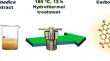
Bio-imaging is a crucial tool for researchers in the fields of cell biology and developmental biomedical sector. Among the various available imaging techniques, fluorescence based imaging stands out due to its high sensitivity and specificity. However, traditional fluorescent materials used in biological imaging often suffer from issues such as photostability and biocompatibility. Moreover, plant tissues contain compounds that cause autofluorescence and light scattering, which can hinder fluorescence microscopy effectiveness. This study explores the development of fluorescent carbon dots (Cm-CDs) synthesized from Citrus medica fruit extract for the fluorescence imaging of Vigna radiata root cells. The successful synthesis of CDs with an average size of 6.7 nm is confirmed by Transmission Electron Microscopy (TEM). The X-ray diffraction (XRD) analysis and raman spectroscopy indicated that the obtained CDs are amorphous in nature. The presence of various functional groups on the surface of CDs were identified by Fourier transform infrared (FTIR) spectra. The optical characteristics of Cm-CDs were studied by UV-Visible spectroscopy and photoluminescence spectroscopy. Cm-CDs demonstrated strong excitation-dependent fluorescence, good solubility, and effective penetration in to the Vigna radiata root cells with multicolor luminescence, and addressed autofluorescence issues. Additionally, a comparative analysis determined the optimal concentration for high-resolution, multi-color root cell imaging, with Cm-CD2 (2.5 mg/ml) exhibiting the highest photoluminescence (PL) intensity. These findings highlight the potential of Cm-CDs in enhancing direct endocytosis and overcoming autofluorescence in plant cell imaging, offering promising advancements for cell biology research.
期刊介绍:
Journal of Fluorescence is an international forum for the publication of peer-reviewed original articles that advance the practice of this established spectroscopic technique. Topics covered include advances in theory/and or data analysis, studies of the photophysics of aromatic molecules, solvent, and environmental effects, development of stationary or time-resolved measurements, advances in fluorescence microscopy, imaging, photobleaching/recovery measurements, and/or phosphorescence for studies of cell biology, chemical biology and the advanced uses of fluorescence in flow cytometry/analysis, immunology, high throughput screening/drug discovery, DNA sequencing/arrays, genomics and proteomics. Typical applications might include studies of macromolecular dynamics and conformation, intracellular chemistry, and gene expression. The journal also publishes papers that describe the synthesis and characterization of new fluorophores, particularly those displaying unique sensitivities and/or optical properties. In addition to original articles, the Journal also publishes reviews, rapid communications, short communications, letters to the editor, topical news articles, and technical and design notes.
 求助内容:
求助内容: 应助结果提醒方式:


