{"title":"Development of a plant-based surgical training model for fluorescence-guided cancer surgery","authors":"Mayu Shigeyama MD, Naoki Nishio MD, PhD, Akihisa Wada MD, PhD, Sohei Mitani MD, PhD, Gaku Morimoto MSc, Sayaka Yokoi MD, PhD, Nobuaki Mukoyama MD, PhD, Mai Yokoi MD, Stan van Keulen MD, DDS, PhD, Eben Rosenthal MD, Michihiko Sone MD, PhD","doi":"10.1002/hed.27835","DOIUrl":null,"url":null,"abstract":"<div>\n \n \n <section>\n \n <h3> Background</h3>\n \n <p>Fluorescence-guided surgery (FGS) can help surgeons to discriminate tumor tissue from adjacent normal tissues using fluorescent tracers.</p>\n </section>\n \n <section>\n \n <h3> Methods</h3>\n \n <p>We developed a surgical training model, manufactured using sustainable vegetable organic material with indocyanine green (ICG)-containing “tumor.” Surgeons evaluated the model with both the closed-field and endoscopic fluorescence imaging devices and assessed its efficacy to identify residual tumor after enucleation using electrocautery.</p>\n </section>\n \n <section>\n \n <h3> Results</h3>\n \n <p>Strong correlations of fluorescence were obtained at all working distance (3, 5, 7, and 10 cm), showing the robustness of fluorescence signal for the closed-field and endoscopic fluorescence imaging devices. The higher fluorescence signals were obtained in the wound bed in the closed-field fluorescence imaging device and the residual tumor could be clearly identified by fluorescence endoscopy.</p>\n </section>\n \n <section>\n \n <h3> Conclusions</h3>\n \n <p>Our FGS training model may provide experience for surgeons unfamiliar with optical surgery and subsequent tissue interactions. The model seemed particularly helpful in teaching surgeons the principles of FGS.</p>\n </section>\n </div>","PeriodicalId":55072,"journal":{"name":"Head and Neck-Journal for the Sciences and Specialties of the Head and Neck","volume":null,"pages":null},"PeriodicalIF":2.3000,"publicationDate":"2024-06-06","publicationTypes":"Journal Article","fieldsOfStudy":null,"isOpenAccess":false,"openAccessPdf":"https://onlinelibrary.wiley.com/doi/epdf/10.1002/hed.27835","citationCount":"0","resultStr":null,"platform":"Semanticscholar","paperid":null,"PeriodicalName":"Head and Neck-Journal for the Sciences and Specialties of the Head and Neck","FirstCategoryId":"3","ListUrlMain":"https://onlinelibrary.wiley.com/doi/10.1002/hed.27835","RegionNum":3,"RegionCategory":"医学","ArticlePicture":[],"TitleCN":null,"AbstractTextCN":null,"PMCID":null,"EPubDate":"","PubModel":"","JCR":"Q1","JCRName":"OTORHINOLARYNGOLOGY","Score":null,"Total":0}
引用次数: 0
Abstract
Background
Fluorescence-guided surgery (FGS) can help surgeons to discriminate tumor tissue from adjacent normal tissues using fluorescent tracers.
Methods
We developed a surgical training model, manufactured using sustainable vegetable organic material with indocyanine green (ICG)-containing “tumor.” Surgeons evaluated the model with both the closed-field and endoscopic fluorescence imaging devices and assessed its efficacy to identify residual tumor after enucleation using electrocautery.
Results
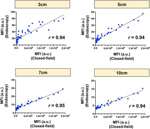
Strong correlations of fluorescence were obtained at all working distance (3, 5, 7, and 10 cm), showing the robustness of fluorescence signal for the closed-field and endoscopic fluorescence imaging devices. The higher fluorescence signals were obtained in the wound bed in the closed-field fluorescence imaging device and the residual tumor could be clearly identified by fluorescence endoscopy.
Conclusions
Our FGS training model may provide experience for surgeons unfamiliar with optical surgery and subsequent tissue interactions. The model seemed particularly helpful in teaching surgeons the principles of FGS.
期刊介绍:
Head & Neck is an international multidisciplinary publication of original contributions concerning the diagnosis and management of diseases of the head and neck. This area involves the overlapping interests and expertise of several surgical and medical specialties, including general surgery, neurosurgery, otolaryngology, plastic surgery, oral surgery, dermatology, ophthalmology, pathology, radiotherapy, medical oncology, and the corresponding basic sciences.
 求助内容:
求助内容: 应助结果提醒方式:


