Huy Quang Phi, Suehyb Ghazi Alkhatib, Scott Bruce Raymond, Omar Aftab Choudhri, Jae Won Song
{"title":"Vessel Wall Imaging in Angiogram-Negative Diffuse Subarachnoid Hemorrhage Reveals a Ruptured Lenticulostriate Aneurysm.","authors":"Huy Quang Phi, Suehyb Ghazi Alkhatib, Scott Bruce Raymond, Omar Aftab Choudhri, Jae Won Song","doi":"10.5469/neuroint.2024.00185","DOIUrl":null,"url":null,"abstract":"<p><p>A patient presented with acute onset headache and subsequent unconsciousness. The neurologic exam showed left-sided myoclonic jerking and right flaccid hemiparalysis. Noncontrast computed tomography revealed diffuse subarachnoid hemorrhage (SAH) with acute hydrocephalus. Initial digital subtraction angiography (DSA) showed no culprit source for SAH. Repeat DSA on day 7 after initial presentation raised suspicion for left internal carotid artery ophthalmic segment and left lateral lenticulostriate artery (LSA) aneurysms. A magnetic resonance vessel wall imaging (VWI) exam was performed given the presence of multiple potential culprit aneurysms. Vessel wall enhancement around the dome of the left LSA aneurysm suggested rupture, which then facilitated treatment with surgical clipping. LSA aneurysms are exceedingly rare and challenging to treat. Given the associated high degree of morbidity, expedient diagnosis is critical to direct management. VWI could be a valuable tool for detecting ruptured aneurysms in the setting of angiogram-negative SAH.</p>","PeriodicalId":19140,"journal":{"name":"Neurointervention","volume":" ","pages":"118-122"},"PeriodicalIF":1.2000,"publicationDate":"2024-07-01","publicationTypes":"Journal Article","fieldsOfStudy":null,"isOpenAccess":false,"openAccessPdf":"https://www.ncbi.nlm.nih.gov/pmc/articles/PMC11222677/pdf/","citationCount":"0","resultStr":null,"platform":"Semanticscholar","paperid":null,"PeriodicalName":"Neurointervention","FirstCategoryId":"1085","ListUrlMain":"https://doi.org/10.5469/neuroint.2024.00185","RegionNum":0,"RegionCategory":null,"ArticlePicture":[],"TitleCN":null,"AbstractTextCN":null,"PMCID":null,"EPubDate":"2024/6/5 0:00:00","PubModel":"Epub","JCR":"Q4","JCRName":"CLINICAL NEUROLOGY","Score":null,"Total":0}
引用次数: 0
Abstract
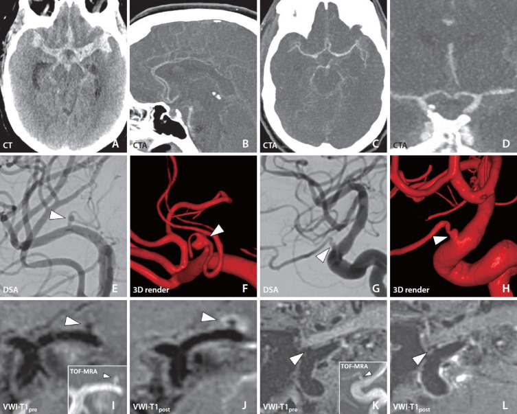
A patient presented with acute onset headache and subsequent unconsciousness. The neurologic exam showed left-sided myoclonic jerking and right flaccid hemiparalysis. Noncontrast computed tomography revealed diffuse subarachnoid hemorrhage (SAH) with acute hydrocephalus. Initial digital subtraction angiography (DSA) showed no culprit source for SAH. Repeat DSA on day 7 after initial presentation raised suspicion for left internal carotid artery ophthalmic segment and left lateral lenticulostriate artery (LSA) aneurysms. A magnetic resonance vessel wall imaging (VWI) exam was performed given the presence of multiple potential culprit aneurysms. Vessel wall enhancement around the dome of the left LSA aneurysm suggested rupture, which then facilitated treatment with surgical clipping. LSA aneurysms are exceedingly rare and challenging to treat. Given the associated high degree of morbidity, expedient diagnosis is critical to direct management. VWI could be a valuable tool for detecting ruptured aneurysms in the setting of angiogram-negative SAH.

 求助内容:
求助内容: 应助结果提醒方式:


