Camila Lopes Vendrami, Nancy A Hammond, David J Escobar, Zachary Zilber, Meaghan Dwyer, Courtney C Moreno, Pardeep K Mittal, Frank H Miller
{"title":"Imaging of pancreatic serous cystadenoma and common imitators.","authors":"Camila Lopes Vendrami, Nancy A Hammond, David J Escobar, Zachary Zilber, Meaghan Dwyer, Courtney C Moreno, Pardeep K Mittal, Frank H Miller","doi":"10.1007/s00261-024-04337-1","DOIUrl":null,"url":null,"abstract":"<p><p>Pancreatic cystic neoplasms are lesions comprised of cystic components that show different biological behaviors, epidemiology, clinical manifestations, imaging features, and malignant potential and management. Benign cystic neoplasms include serous cystic neoplasms (SCAs). Other pancreatic cystic lesions have malignant potential, such as intraductal papillary mucinous neoplasms and mucinous cystic neoplasms. SCAs can be divided into microcystic (classic appearance), honeycomb, oligocystic/macrocystic, and solid patterns based on imaging appearance. They are usually solitary but may be multiple in von Hippel-Lindau disease, which may depict disseminated involvement. The variable appearances of SCAs can mimic other types of pancreatic cystic lesions, and cross-sectional imaging plays an important role in their differential diagnosis. Endoscopic ultrasonography has helped in improving diagnostic accuracy of pancreatic cystic lesions by guiding tissue sampling (biopsy) or cyst fluid analysis. Immunohistochemistry and newer techniques such as radiomics have shown improved performance for preoperatively discriminating SCAs and their mimickers.</p>","PeriodicalId":7126,"journal":{"name":"Abdominal Radiology","volume":null,"pages":null},"PeriodicalIF":2.3000,"publicationDate":"2024-10-01","publicationTypes":"Journal Article","fieldsOfStudy":null,"isOpenAccess":false,"openAccessPdf":"","citationCount":"0","resultStr":null,"platform":"Semanticscholar","paperid":null,"PeriodicalName":"Abdominal Radiology","FirstCategoryId":"3","ListUrlMain":"https://doi.org/10.1007/s00261-024-04337-1","RegionNum":3,"RegionCategory":"医学","ArticlePicture":[],"TitleCN":null,"AbstractTextCN":null,"PMCID":null,"EPubDate":"2024/6/2 0:00:00","PubModel":"Epub","JCR":"Q2","JCRName":"RADIOLOGY, NUCLEAR MEDICINE & MEDICAL IMAGING","Score":null,"Total":0}
引用次数: 0
Abstract
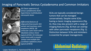
Pancreatic cystic neoplasms are lesions comprised of cystic components that show different biological behaviors, epidemiology, clinical manifestations, imaging features, and malignant potential and management. Benign cystic neoplasms include serous cystic neoplasms (SCAs). Other pancreatic cystic lesions have malignant potential, such as intraductal papillary mucinous neoplasms and mucinous cystic neoplasms. SCAs can be divided into microcystic (classic appearance), honeycomb, oligocystic/macrocystic, and solid patterns based on imaging appearance. They are usually solitary but may be multiple in von Hippel-Lindau disease, which may depict disseminated involvement. The variable appearances of SCAs can mimic other types of pancreatic cystic lesions, and cross-sectional imaging plays an important role in their differential diagnosis. Endoscopic ultrasonography has helped in improving diagnostic accuracy of pancreatic cystic lesions by guiding tissue sampling (biopsy) or cyst fluid analysis. Immunohistochemistry and newer techniques such as radiomics have shown improved performance for preoperatively discriminating SCAs and their mimickers.
胰腺囊性肿瘤是由囊性成分组成的病变,它们表现出不同的生物学行为、流行病学、临床表现、影像学特征以及恶性潜能和治疗方法。良性囊性瘤包括浆液性囊性瘤(SCA)。其他胰腺囊性病变具有恶性潜能,如导管内乳头状粘液瘤和粘液性囊性瘤。根据影像学表现,胰腺囊肿可分为微囊型(典型表现)、蜂窝型、少囊型/巨囊型和实性型。它们通常是单发的,但在 von Hippel-Lindau 病中可能是多发的,这可能描述了播散性受累。SCA的外观多变,可模仿其他类型的胰腺囊性病变,因此横断面成像在其鉴别诊断中起着重要作用。内镜超声检查通过引导组织取样(活检)或囊液分析,有助于提高胰腺囊性病变的诊断准确性。免疫组化和放射组学等新技术在术前鉴别胰腺囊肿及其模仿者方面的性能有所提高。
期刊介绍:
Abdominal Radiology seeks to meet the professional needs of the abdominal radiologist by publishing clinically pertinent original, review and practice related articles on the gastrointestinal and genitourinary tracts and abdominal interventional and radiologic procedures. Case reports are generally not accepted unless they are the first report of a new disease or condition, or part of a special solicited section.
Reasons to Publish Your Article in Abdominal Radiology:
· Official journal of the Society of Abdominal Radiology (SAR)
· Published in Cooperation with:
European Society of Gastrointestinal and Abdominal Radiology (ESGAR)
European Society of Urogenital Radiology (ESUR)
Asian Society of Abdominal Radiology (ASAR)
· Efficient handling and Expeditious review
· Author feedback is provided in a mentoring style
· Global readership
· Readers can earn CME credits

 求助内容:
求助内容: 应助结果提醒方式:


