Comparative macroanatomical and scanning electron microscopy study of the eyeball in brachycephalic and mesocephalic dog breeds
Abstract
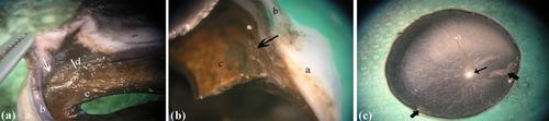
The anatomical structures forming the eyeball differ among dog breeds, both morphologically and morphometrically. This study was aimed at determining the morphometric values of the eyeball layers of different dog breeds and the morphological structures of these layers using scanning electron microscopy. Thirty-two eyeballs of 17 dogs belonging to 9 different breeds that died from traffic accidents, falling from a height, and naturally were used. These dog breeds were grouped according to their brachycephalic and mesocephalic skull structures, and morphometric measurements of the eyeballs of each group were obtained. Scanning electron microscopy was used to examine the morphological structure of the eyeball layers. The studied dogs' eyeballs comprised three layers: outer, middle, and inner. Thickness measurements obtained from three different regions of the eyeball indicated that the equatorial region was the thinnest among all dog breeds. Moreover, the cornea, which is covered by the sclera along its edges, was thicker at the corneal limbus than at the corneal vertex. A positive correlation was observed between lens thickness and the number of ciliary processes, which varied according to the dogs' head structures. Notably, depression was observed in the posterior surface of the lens in brachycephalic dogs. The morphometric values of the eyeball layers in the brachycephalic and mesocephalic dog breeds were also determined. These values will help researchers study this subject, and the determined morphometric and morphological values will contribute to the anatomy literature.
Research Highlights
- This comprehensive study investigates the morphometric and morphological variations in the eyeball layers of different dog breeds, utilizing scanning electron microscopy to analyze eyeballs. It reveals significant breed-specific differences, particularly between brachycephalic and mesocephalic dogs, regarding eyeball layer thickness, corneal structure, lens thickness, and the number of ciliary processes.

 求助内容:
求助内容: 应助结果提醒方式:


