Wen-Wen Kong, Yun Zhu, Heng-Rui Zhao, Kang Du, Rui-Qian Zhou, Bo Li, Feng Yang, Pu Hou, Xia-He Huang, Yuxing Chen, Ying-Chun Wang, Fei Sun, Yong-Liang Jiang, Cong-Zhao Zhou
{"title":"Cryo-electron tomography reveals the packaging pattern of RuBisCOs in Synechococcus β-carboxysome","authors":"Wen-Wen Kong, Yun Zhu, Heng-Rui Zhao, Kang Du, Rui-Qian Zhou, Bo Li, Feng Yang, Pu Hou, Xia-He Huang, Yuxing Chen, Ying-Chun Wang, Fei Sun, Yong-Liang Jiang, Cong-Zhao Zhou","doi":"10.1016/j.str.2024.05.007","DOIUrl":null,"url":null,"abstract":"<p>Carboxysomes are large self-assembled microcompartments that serve as the central machinery of a CO<sub>2</sub>-concentrating mechanism (CCM). Biogenesis of carboxysome requires the fine organization of thousands of individual proteins; however, the packaging pattern of internal RuBisCOs remains largely unknown. Here we purified the intact β-carboxysomes from <em>Synechococcus elongatus</em> PCC 7942 and identified the protein components by mass spectrometry. Cryo-electron tomography combined with subtomogram averaging revealed the general organization pattern of internal RuBisCOs, in which the adjacent RuBisCOs are mainly arranged in three distinct manners: head-to-head, head-to-side, and side-by-side. The RuBisCOs in the outermost layer are regularly aligned along the shell, the majority of which directly interact with the shell. Moreover, statistical analysis enabled us to propose an ideal packaging model of RuBisCOs in the β-carboxysome. These results provide new insights into the biogenesis of β-carboxysomes and also advance our understanding of the efficient carbon fixation functionality of carboxysomes.</p>","PeriodicalId":22168,"journal":{"name":"Structure","volume":null,"pages":null},"PeriodicalIF":4.4000,"publicationDate":"2024-05-31","publicationTypes":"Journal Article","fieldsOfStudy":null,"isOpenAccess":false,"openAccessPdf":"","citationCount":"0","resultStr":null,"platform":"Semanticscholar","paperid":null,"PeriodicalName":"Structure","FirstCategoryId":"99","ListUrlMain":"https://doi.org/10.1016/j.str.2024.05.007","RegionNum":2,"RegionCategory":"生物学","ArticlePicture":[],"TitleCN":null,"AbstractTextCN":null,"PMCID":null,"EPubDate":"","PubModel":"","JCR":"Q2","JCRName":"BIOCHEMISTRY & MOLECULAR BIOLOGY","Score":null,"Total":0}
引用次数: 0
Abstract
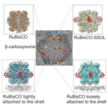
Carboxysomes are large self-assembled microcompartments that serve as the central machinery of a CO2-concentrating mechanism (CCM). Biogenesis of carboxysome requires the fine organization of thousands of individual proteins; however, the packaging pattern of internal RuBisCOs remains largely unknown. Here we purified the intact β-carboxysomes from Synechococcus elongatus PCC 7942 and identified the protein components by mass spectrometry. Cryo-electron tomography combined with subtomogram averaging revealed the general organization pattern of internal RuBisCOs, in which the adjacent RuBisCOs are mainly arranged in three distinct manners: head-to-head, head-to-side, and side-by-side. The RuBisCOs in the outermost layer are regularly aligned along the shell, the majority of which directly interact with the shell. Moreover, statistical analysis enabled us to propose an ideal packaging model of RuBisCOs in the β-carboxysome. These results provide new insights into the biogenesis of β-carboxysomes and also advance our understanding of the efficient carbon fixation functionality of carboxysomes.
期刊介绍:
Structure aims to publish papers of exceptional interest in the field of structural biology. The journal strives to be essential reading for structural biologists, as well as biologists and biochemists that are interested in macromolecular structure and function. Structure strongly encourages the submission of manuscripts that present structural and molecular insights into biological function and mechanism. Other reports that address fundamental questions in structural biology, such as structure-based examinations of protein evolution, folding, and/or design, will also be considered. We will consider the application of any method, experimental or computational, at high or low resolution, to conduct structural investigations, as long as the method is appropriate for the biological, functional, and mechanistic question(s) being addressed. Likewise, reports describing single-molecule analysis of biological mechanisms are welcome.
In general, the editors encourage submission of experimental structural studies that are enriched by an analysis of structure-activity relationships and will not consider studies that solely report structural information unless the structure or analysis is of exceptional and broad interest. Studies reporting only homology models, de novo models, or molecular dynamics simulations are also discouraged unless the models are informed by or validated by novel experimental data; rationalization of a large body of existing experimental evidence and making testable predictions based on a model or simulation is often not considered sufficient.
 求助内容:
求助内容: 应助结果提醒方式:


