Ashok Kumar Maurya, Ashish Kumar Pandey, Piyali Deb Barman
{"title":"Chromite ore: X-ray fluorescence spectral analysis of Kβ and L-lines by WD-XRF","authors":"Ashok Kumar Maurya, Ashish Kumar Pandey, Piyali Deb Barman","doi":"10.1007/s12039-024-02280-8","DOIUrl":null,"url":null,"abstract":"<div><p>This work delves into the utility of chromium’s X-ray fluorescence (XRF) lines for the analysis of chromite ores, employing a wavelength-dispersive XRF spectrometer. The CrL<i>α</i><sub>1,2</sub> and CrL<i>β</i><sub>1</sub> fluorescence lines, despite not involving electronic transitions from valence to K-core shell, offer substantial insights into the valence state of chromium. This study examines these relatively unexplored but potentially valuable lines and compares them with the CrK<i>β</i> series fluorescence lines. Intriguingly, our investigation reveals striking differences in the L-line fluorescence spectra between metallic chromium (Cr(0)), chromite ores (Fe, Mg)Cr<sub>2</sub>O<sub>4</sub>, and K<sub>2</sub>Cr<sub>2</sub>O<sub>7</sub>. Chemical shifts and peak shapes emerge as key discriminators, surpassing the sensitivity of the CrK<i>β</i><sub>1,3</sub> lines. Notably, the chemical shifts of L-lines are amplified, showcasing a clear trend: Cr(III) exhibits a larger shift than Cr(VI), mirroring the pattern observed in the K<i>β</i>-lines. The fluorescence peak shapes of L<i>α</i><sub>1,2</sub> and L<i>β</i><sub>1</sub> for metallic chromium and chromite ores (both have unpaired 3d-electrons) are similar, whereas those of K<sub>2</sub>Cr<sub>2</sub>O<sub>7</sub> differ considerably in peak shape, indicating that unpaired 3d-electrons influence these peaks. A linear relationship was found between the area under the curve ratio for L<i>α</i><sub>1,2</sub> and L<i>β</i><sub>1</sub> and the valence state of chromium.</p><h3>Graphical Abstract</h3>\n<div><figure><div><div><picture><source><img></source></picture></div></div></figure></div></div>","PeriodicalId":616,"journal":{"name":"Journal of Chemical Sciences","volume":"136 2","pages":""},"PeriodicalIF":1.7000,"publicationDate":"2024-05-28","publicationTypes":"Journal Article","fieldsOfStudy":null,"isOpenAccess":false,"openAccessPdf":"https://link.springer.com/content/pdf/10.1007/s12039-024-02280-8.pdf","citationCount":"0","resultStr":null,"platform":"Semanticscholar","paperid":null,"PeriodicalName":"Journal of Chemical Sciences","FirstCategoryId":"92","ListUrlMain":"https://link.springer.com/article/10.1007/s12039-024-02280-8","RegionNum":4,"RegionCategory":"化学","ArticlePicture":[],"TitleCN":null,"AbstractTextCN":null,"PMCID":null,"EPubDate":"","PubModel":"","JCR":"Q3","JCRName":"CHEMISTRY, MULTIDISCIPLINARY","Score":null,"Total":0}
引用次数: 0
Abstract
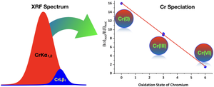
This work delves into the utility of chromium’s X-ray fluorescence (XRF) lines for the analysis of chromite ores, employing a wavelength-dispersive XRF spectrometer. The CrLα1,2 and CrLβ1 fluorescence lines, despite not involving electronic transitions from valence to K-core shell, offer substantial insights into the valence state of chromium. This study examines these relatively unexplored but potentially valuable lines and compares them with the CrKβ series fluorescence lines. Intriguingly, our investigation reveals striking differences in the L-line fluorescence spectra between metallic chromium (Cr(0)), chromite ores (Fe, Mg)Cr2O4, and K2Cr2O7. Chemical shifts and peak shapes emerge as key discriminators, surpassing the sensitivity of the CrKβ1,3 lines. Notably, the chemical shifts of L-lines are amplified, showcasing a clear trend: Cr(III) exhibits a larger shift than Cr(VI), mirroring the pattern observed in the Kβ-lines. The fluorescence peak shapes of Lα1,2 and Lβ1 for metallic chromium and chromite ores (both have unpaired 3d-electrons) are similar, whereas those of K2Cr2O7 differ considerably in peak shape, indicating that unpaired 3d-electrons influence these peaks. A linear relationship was found between the area under the curve ratio for Lα1,2 and Lβ1 and the valence state of chromium.
期刊介绍:
Journal of Chemical Sciences is a monthly journal published by the Indian Academy of Sciences. It formed part of the original Proceedings of the Indian Academy of Sciences – Part A, started by the Nobel Laureate Prof C V Raman in 1934, that was split in 1978 into three separate journals. It was renamed as Journal of Chemical Sciences in 2004. The journal publishes original research articles and rapid communications, covering all areas of chemical sciences. A significant feature of the journal is its special issues, brought out from time to time, devoted to conference symposia/proceedings in frontier areas of the subject, held not only in India but also in other countries.
 求助内容:
求助内容: 应助结果提醒方式:


