Macroscopic, microscopic, and immunofluorescent characterization of the Greek tortoise (Testudo graeca graeca) oropharyngeal floor with concern to its feed adaptation as a herbivorous land reptile
Abstract
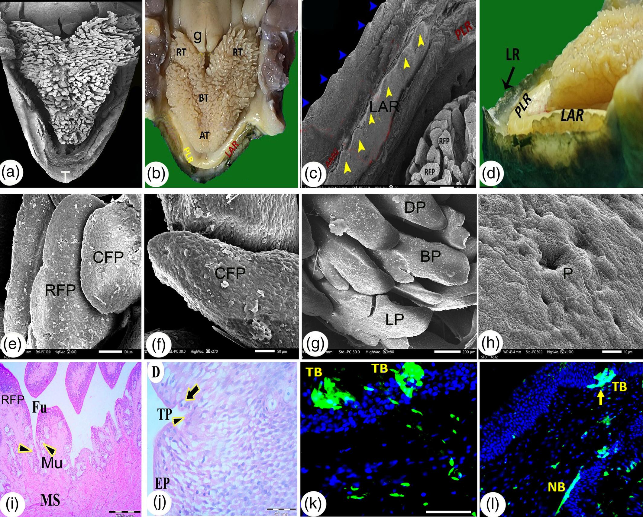
The current investigation focuses on gross anatomy, light, and scanning electron microscopy (SEM) of the Testudo graeca oropharyngeal floor, with particular reference to the immunofluorescence technique to examine its tongue. The T. graeca oropharyngeal floor showed many anatomical structures: the lower rhamphotheca, paralingual ridge, lower alveolar ridge, tongue, laryngeal mound, and glottis. The lower rhamphotheca appeared as a V-shaped jaw line with a highly serrated edge and a median tomium (beak). SEM observations of the lingual apex and the lingual body showed rectangular and conical filiform papillae with porous surfaces and taste pores. Meanwhile, the lingual root had two wings that carried papillae with different shapes: dagger-shaped, conical, bifurcated, and leaf-like papillae, and these papillae lacked taste pores. The laryngeal mound had openings for the laryngeal mucus gland and its secretions. Light microscopy findings showed mucous glands in the propria submucosa and near the mucosal surface of the lingual apex. The lingual root had lingual papillae and two hyaline cartilaginous skeletons between skeletal muscles, and the lingual papillae were elongated filiform, rectangular filiform papillae, and fungiform papillae. The lamina propria constituted the core of the lingual papillae and the mucous gland, they had a positive reaction with the periodic acid schiff (PAS) reagent. The apical surface of the fungiform papillae had taste pores. Under immunofluorescence, the vimentin was detected in taste bud cells, and synaptophysin reacted to the taste buds and nerve bundles. The current study of the Greek tortoise oropharyngeal floor investigated its herbivorous eating habits using its serrated lower rhamphotheca, a large tongue with differently shaped papillae, and numerous mucous glands.
Research Highlights
- The Greek tortoise (T. graeca graeca) oropharyngeal floor showed many anatomical structures: lower rhamphotheca, paralingual ridge, lower alveolar ridge, tongue, laryngeal mound, and glottis.
- SEM and light microscopy observations of the tongue revealed varied types and shapes of lingual papillae with a porous surface on the tongue apex (rectangular or conical filiform papillae), on the tongue body (filiform and fungiform papillae), and on the tongue root (dagger-shaped, conical, bifurcated, and leaf-like papillae).
- Light microscopy findings: the lamina propria constituted the core of the lingual papillae and had numerous mucous glands that had a slightly magenta-red color with PAS reagent.
- The apical surface of the fungiform papillae had taste pores. Vimentin and synaptophysin gave a reaction to the taste buds.

 求助内容:
求助内容: 应助结果提醒方式:


