{"title":"Optimising flow without congestion using the venous-arterial Doppler enhanced resuscitation framework","authors":"Jon-Emile S Kenny, Philippe Rola","doi":"10.1002/ajum.12388","DOIUrl":null,"url":null,"abstract":"<div>\n \n \n <section>\n \n <h3> Introduction</h3>\n \n <p>Ultrasonography as a guide for intravenous (IV) fluid therapy is increasingly accepted within the spheres of acute care. Initial investigations and protocols often focused on measures of arterial flow as an objective approach for personalising organ ‘perfusion.’ More recently, and with literature associating excessive IV fluid with adverse outcomes, venous ultrasound as a measure of organ ‘congestion’ is taking hold. Yet, arterial (i.e., ‘perfusion’) and venous (i.e., ‘congestion’) Doppler ultrasound measures are often performed separately and can be time-consuming, especially for novices.</p>\n </section>\n \n <section>\n \n <h3> Methods</h3>\n \n <p>We report a case, wherein venous and arterial Doppler were simultaneously measured using a wireless, wearable ultrasound as a means to optimise flow without congestion.</p>\n </section>\n \n <section>\n \n <h3> Results</h3>\n \n <p>Before IV volume expansion, the patient had Doppler measures consistent with low central venous pressure (CVP) and stroke volume (SV). Following IV volume expansion, venous Doppler remained the same; however, carotid corrected flow time (ccFT) increased significantly.</p>\n </section>\n \n <section>\n \n <h3> Conclusion</h3>\n \n <p>A framework for venous-arterial Doppler enhanced resuscitation (VADER) can be used to guide IV volume in patients at risk for venous congestion.</p>\n </section>\n </div>","PeriodicalId":36517,"journal":{"name":"Australasian Journal of Ultrasound in Medicine","volume":"27 3","pages":"193-196"},"PeriodicalIF":0.0000,"publicationDate":"2024-05-08","publicationTypes":"Journal Article","fieldsOfStudy":null,"isOpenAccess":false,"openAccessPdf":"https://onlinelibrary.wiley.com/doi/epdf/10.1002/ajum.12388","citationCount":"0","resultStr":null,"platform":"Semanticscholar","paperid":null,"PeriodicalName":"Australasian Journal of Ultrasound in Medicine","FirstCategoryId":"1085","ListUrlMain":"https://onlinelibrary.wiley.com/doi/10.1002/ajum.12388","RegionNum":0,"RegionCategory":null,"ArticlePicture":[],"TitleCN":null,"AbstractTextCN":null,"PMCID":null,"EPubDate":"","PubModel":"","JCR":"Q3","JCRName":"Medicine","Score":null,"Total":0}
引用次数: 0
Abstract
Introduction
Ultrasonography as a guide for intravenous (IV) fluid therapy is increasingly accepted within the spheres of acute care. Initial investigations and protocols often focused on measures of arterial flow as an objective approach for personalising organ ‘perfusion.’ More recently, and with literature associating excessive IV fluid with adverse outcomes, venous ultrasound as a measure of organ ‘congestion’ is taking hold. Yet, arterial (i.e., ‘perfusion’) and venous (i.e., ‘congestion’) Doppler ultrasound measures are often performed separately and can be time-consuming, especially for novices.
Methods
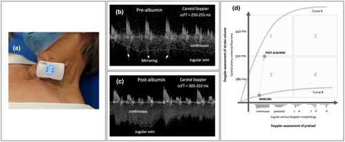
We report a case, wherein venous and arterial Doppler were simultaneously measured using a wireless, wearable ultrasound as a means to optimise flow without congestion.
Results
Before IV volume expansion, the patient had Doppler measures consistent with low central venous pressure (CVP) and stroke volume (SV). Following IV volume expansion, venous Doppler remained the same; however, carotid corrected flow time (ccFT) increased significantly.
Conclusion
A framework for venous-arterial Doppler enhanced resuscitation (VADER) can be used to guide IV volume in patients at risk for venous congestion.

 求助内容:
求助内容: 应助结果提醒方式:


