Neurochemistry and circuit organization of the lateral spiriform nucleus of birds: A uniquely nonmammalian direct pathway component of the basal ganglia
Anton Reiner, Loreta Medina, Antonio Abellan, Yunping Deng, Claudio A. B. Toledo, Harald Luksch, Tomas Vega-Zuniga, Nell B. Riley, William Hodos, Harvey J. Karten
{"title":"Neurochemistry and circuit organization of the lateral spiriform nucleus of birds: A uniquely nonmammalian direct pathway component of the basal ganglia","authors":"Anton Reiner, Loreta Medina, Antonio Abellan, Yunping Deng, Claudio A. B. Toledo, Harald Luksch, Tomas Vega-Zuniga, Nell B. Riley, William Hodos, Harvey J. Karten","doi":"10.1002/cne.25620","DOIUrl":null,"url":null,"abstract":"<p>We used diverse methods to characterize the role of avian lateral spiriform nucleus (SpL) in basal ganglia motor function. Connectivity analysis showed that SpL receives input from globus pallidus (GP), and the intrapeduncular nucleus (INP) located ventromedial to GP, whose neurons express numerous striatal markers. SpL-projecting GP neurons were large and aspiny, while SpL-projecting INP neurons were medium sized and spiny. Connectivity analysis further showed that SpL receives inputs from subthalamic nucleus (STN) and substantia nigra pars reticulata (SNr), and that the SNr also receives inputs from GP, INP, and STN. Neurochemical analysis showed that SpL neurons express ENK, GAD, and a variety of pallidal neuron markers, and receive GABAergic terminals, some of which also contain DARPP32, consistent with GP pallidal and INP striatal inputs. Connectivity and neurochemical analysis showed that the SpL input to tectum prominently ends on GABAA receptor-enriched tectobulbar neurons. Behavioral studies showed that lesions of SpL impair visuomotor behaviors involving tracking and pecking moving targets. Our results suggest that SpL modulates brainstem-projecting tectobulbar neurons in a manner comparable to the demonstrated influence of GP internus on motor thalamus and of SNr on tectobulbar neurons in mammals. Given published data in amphibians and reptiles, it seems likely the SpL circuit represents a major direct pathway-type circuit by which the basal ganglia exerts its motor influence in nonmammalian tetrapods. The present studies also show that avian striatum is divided into three spatially segregated territories with differing connectivity, a medial striato-nigral territory, a dorsolateral striato-GP territory, and the ventrolateral INP motor territory.</p>","PeriodicalId":15552,"journal":{"name":"Journal of Comparative Neurology","volume":"532 5","pages":""},"PeriodicalIF":2.3000,"publicationDate":"2024-05-11","publicationTypes":"Journal Article","fieldsOfStudy":null,"isOpenAccess":false,"openAccessPdf":"","citationCount":"0","resultStr":null,"platform":"Semanticscholar","paperid":null,"PeriodicalName":"Journal of Comparative Neurology","FirstCategoryId":"3","ListUrlMain":"https://onlinelibrary.wiley.com/doi/10.1002/cne.25620","RegionNum":4,"RegionCategory":"医学","ArticlePicture":[],"TitleCN":null,"AbstractTextCN":null,"PMCID":null,"EPubDate":"","PubModel":"","JCR":"Q3","JCRName":"NEUROSCIENCES","Score":null,"Total":0}
引用次数: 0
Abstract
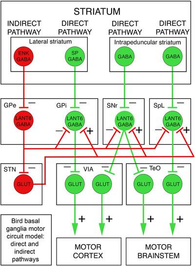
We used diverse methods to characterize the role of avian lateral spiriform nucleus (SpL) in basal ganglia motor function. Connectivity analysis showed that SpL receives input from globus pallidus (GP), and the intrapeduncular nucleus (INP) located ventromedial to GP, whose neurons express numerous striatal markers. SpL-projecting GP neurons were large and aspiny, while SpL-projecting INP neurons were medium sized and spiny. Connectivity analysis further showed that SpL receives inputs from subthalamic nucleus (STN) and substantia nigra pars reticulata (SNr), and that the SNr also receives inputs from GP, INP, and STN. Neurochemical analysis showed that SpL neurons express ENK, GAD, and a variety of pallidal neuron markers, and receive GABAergic terminals, some of which also contain DARPP32, consistent with GP pallidal and INP striatal inputs. Connectivity and neurochemical analysis showed that the SpL input to tectum prominently ends on GABAA receptor-enriched tectobulbar neurons. Behavioral studies showed that lesions of SpL impair visuomotor behaviors involving tracking and pecking moving targets. Our results suggest that SpL modulates brainstem-projecting tectobulbar neurons in a manner comparable to the demonstrated influence of GP internus on motor thalamus and of SNr on tectobulbar neurons in mammals. Given published data in amphibians and reptiles, it seems likely the SpL circuit represents a major direct pathway-type circuit by which the basal ganglia exerts its motor influence in nonmammalian tetrapods. The present studies also show that avian striatum is divided into three spatially segregated territories with differing connectivity, a medial striato-nigral territory, a dorsolateral striato-GP territory, and the ventrolateral INP motor territory.
期刊介绍:
Established in 1891, JCN is the oldest continually published basic neuroscience journal. Historically, as the name suggests, the journal focused on a comparison among species to uncover the intricacies of how the brain functions. In modern times, this research is called systems neuroscience where animal models are used to mimic core cognitive processes with the ultimate goal of understanding neural circuits and connections that give rise to behavioral patterns and different neural states.
Research published in JCN covers all species from invertebrates to humans, and the reports inform the readers about the function and organization of nervous systems in species with an emphasis on the way that species adaptations inform about the function or organization of the nervous systems, rather than on their evolution per se.
JCN publishes primary research articles and critical commentaries and review-type articles offering expert insight in to cutting edge research in the field of systems neuroscience; a complete list of contribution types is given in the Author Guidelines. For primary research contributions, only full-length investigative reports are desired; the journal does not accept short communications.

 求助内容:
求助内容: 应助结果提醒方式:


