Syed Nasar Shah, Sezai Ercisli, Akhtar Zaman, Israr Ud Din, Fahim Ullah Khan, Muhammad Nauman Khan, Majid Iqbal, Baber Ali, Mohammad K. Okla
{"title":"SEM and LM insights: Spore morphology and its taxonomic significance in Sino Himalayan, Malesian, and European elements of Pteridaceae","authors":"Syed Nasar Shah, Sezai Ercisli, Akhtar Zaman, Israr Ud Din, Fahim Ullah Khan, Muhammad Nauman Khan, Majid Iqbal, Baber Ali, Mohammad K. Okla","doi":"10.1002/jemt.24583","DOIUrl":null,"url":null,"abstract":"<div>\n \n \n <section>\n \n <p>The Pteridaceae family, known for its taxonomic complexity, presents challenges in identification due to high variability among its species. This study investigates the spore morphology employing both SEM and LM techniques in 10 Pteridaceae taxa phytogeographicaly Sino-Himalayan, Malesian, and European elements in Pakistan. The taxa include <i>Adiantum capillus-veneris</i>, <i>A. incisum</i>, <i>A. venustum</i>, <i>Aleuritopteris bicolor</i>, <i>Oeosporangium nitidulum</i>, <i>O. pteridioides</i>, <i>Onychium cryptogrammoides</i>, <i>O. vermae</i>, <i>Pteris cretica</i>, and <i>P. vittata</i>. The objective is to assess their taxonomic relevance and develop a spore-based taxonomic key. Findings indicate differences in spore shape, sizes, exospore thickness, and in surface ornamentation highlighting the potential for taxonomic differentiation. Spores are trilete, and notable differences are observed in the dimension of spores in both distal and proximal sides. Equatorial dimensions vary between 35 and 50 μm, while the polar diameter ranges from 29 to 50 μm. SEM revealed different spore ornamentation types that show several useful characteristics establishing valuable taxonomic variations. The studied <i>Adiantum</i> taxa feature a perispore with tubercules and a micro-granulose surface. The spores of examined <i>Oeosporangium</i> and <i>Aleuritopteris</i> taxa shows cristate sculptures with variable ornamentations. Both species of <i>Onychium</i> have tuberculate-pleated tubercles with sinuous folds on both distal and proximal sides. The surface ornamentation among examined <i>Pteris</i> taxa show variability. PCA analysis indicated that spore quantitative data identified distinct groups, underscoring taxonomic significance. Nevertheless, there was variation observed in surface ornamentation and spore shape, indicating the potential for discrimination among taxa.</p>\n </section>\n \n <section>\n \n <h3> Research Highlights</h3>\n \n <div>\n <ul>\n \n <li>Spore morphology of 10 Pteridaceae taxa has been investigated through LM and SEM.</li>\n \n <li>Investigated species shows differences in spore shape, sizes, exospore thickness, and in surface ornamentation.</li>\n \n <li>Ornamentation on the perispore provides several valuable characteristics, establishing useful taxonomic distinctions.</li>\n \n <li>Spore morphological analysis is effective at the generic level, with minor distinctions discernible at the species level.</li>\n </ul>\n </div>\n </section>\n </div>","PeriodicalId":18684,"journal":{"name":"Microscopy Research and Technique","volume":"87 10","pages":"2253-2265"},"PeriodicalIF":2.0000,"publicationDate":"2024-05-10","publicationTypes":"Journal Article","fieldsOfStudy":null,"isOpenAccess":false,"openAccessPdf":"","citationCount":"0","resultStr":null,"platform":"Semanticscholar","paperid":null,"PeriodicalName":"Microscopy Research and Technique","FirstCategoryId":"5","ListUrlMain":"https://onlinelibrary.wiley.com/doi/10.1002/jemt.24583","RegionNum":3,"RegionCategory":"工程技术","ArticlePicture":[],"TitleCN":null,"AbstractTextCN":null,"PMCID":null,"EPubDate":"","PubModel":"","JCR":"Q2","JCRName":"ANATOMY & MORPHOLOGY","Score":null,"Total":0}
引用次数: 0
Abstract
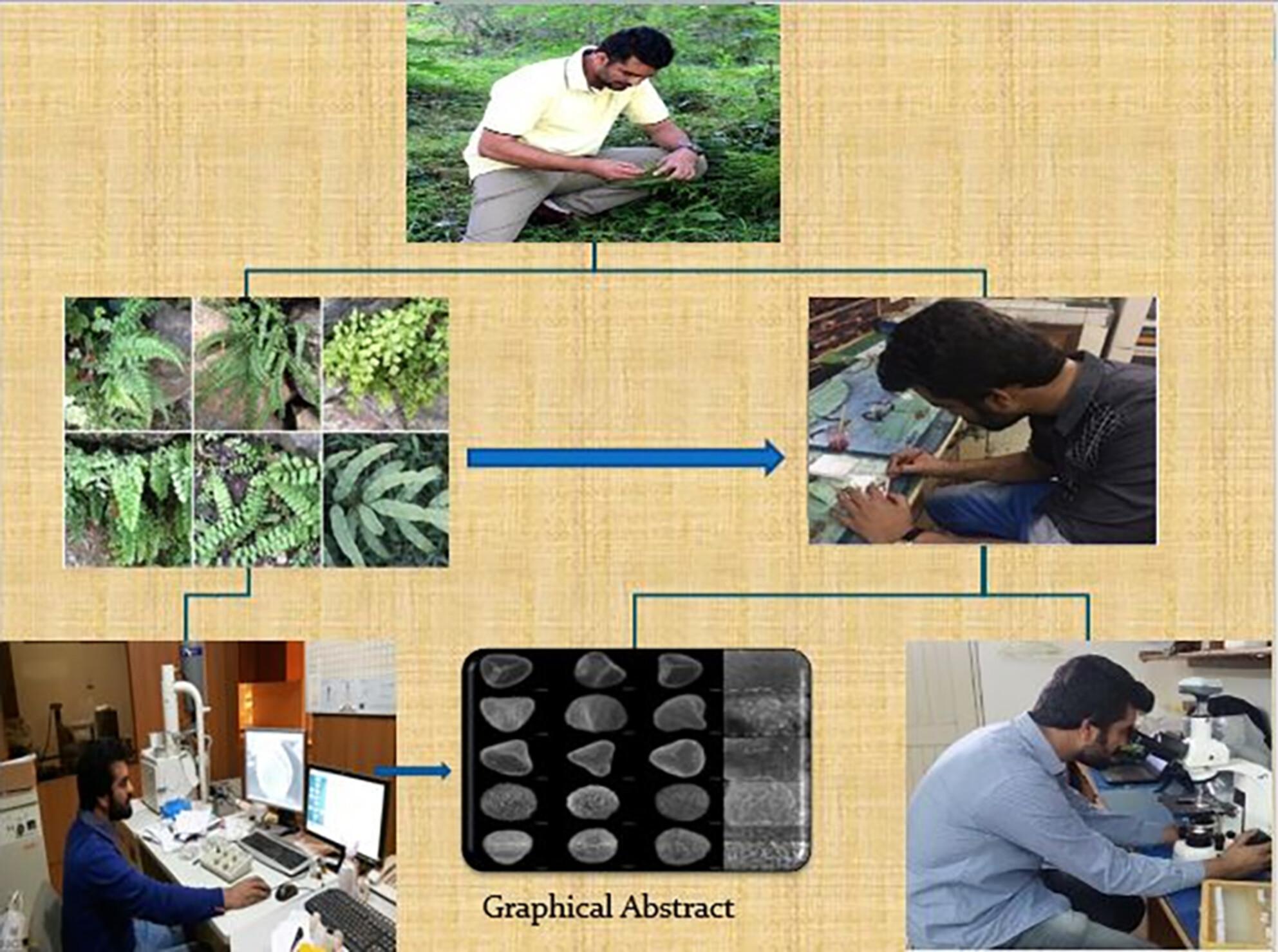
The Pteridaceae family, known for its taxonomic complexity, presents challenges in identification due to high variability among its species. This study investigates the spore morphology employing both SEM and LM techniques in 10 Pteridaceae taxa phytogeographicaly Sino-Himalayan, Malesian, and European elements in Pakistan. The taxa include Adiantum capillus-veneris, A. incisum, A. venustum, Aleuritopteris bicolor, Oeosporangium nitidulum, O. pteridioides, Onychium cryptogrammoides, O. vermae, Pteris cretica, and P. vittata. The objective is to assess their taxonomic relevance and develop a spore-based taxonomic key. Findings indicate differences in spore shape, sizes, exospore thickness, and in surface ornamentation highlighting the potential for taxonomic differentiation. Spores are trilete, and notable differences are observed in the dimension of spores in both distal and proximal sides. Equatorial dimensions vary between 35 and 50 μm, while the polar diameter ranges from 29 to 50 μm. SEM revealed different spore ornamentation types that show several useful characteristics establishing valuable taxonomic variations. The studied Adiantum taxa feature a perispore with tubercules and a micro-granulose surface. The spores of examined Oeosporangium and Aleuritopteris taxa shows cristate sculptures with variable ornamentations. Both species of Onychium have tuberculate-pleated tubercles with sinuous folds on both distal and proximal sides. The surface ornamentation among examined Pteris taxa show variability. PCA analysis indicated that spore quantitative data identified distinct groups, underscoring taxonomic significance. Nevertheless, there was variation observed in surface ornamentation and spore shape, indicating the potential for discrimination among taxa.
Research Highlights
Spore morphology of 10 Pteridaceae taxa has been investigated through LM and SEM.
Investigated species shows differences in spore shape, sizes, exospore thickness, and in surface ornamentation.
Ornamentation on the perispore provides several valuable characteristics, establishing useful taxonomic distinctions.
Spore morphological analysis is effective at the generic level, with minor distinctions discernible at the species level.
期刊介绍:
Microscopy Research and Technique (MRT) publishes articles on all aspects of advanced microscopy original architecture and methodologies with applications in the biological, clinical, chemical, and materials sciences. Original basic and applied research as well as technical papers dealing with the various subsets of microscopy are encouraged. MRT is the right form for those developing new microscopy methods or using the microscope to answer key questions in basic and applied research.