{"title":"Zooming into lipid droplet biology through the lens of electron microscopy","authors":"Wioleta Dudka, Veijo T. Salo, Julia Mahamid","doi":"10.1002/1873-3468.14899","DOIUrl":null,"url":null,"abstract":"<p>Electron microscopy (EM), in its various flavors, has significantly contributed to our understanding of lipid droplets (LD) as central organelles in cellular metabolism. For example, EM has illuminated that LDs, in contrast to all other cellular organelles, are uniquely enclosed by a single phospholipid monolayer, revealed the architecture of LD contact sites with different organelles, and provided near-atomic resolution maps of key enzymes that regulate neutral lipid biosynthesis and LD biogenesis. In this review, we first provide a brief history of pivotal findings in LD biology unveiled through the lens of an electron microscope. We describe the main EM techniques used in the context of LD research and discuss their current capabilities and limitations, thereby providing a foundation for utilizing suitable EM methodology to address LD-related questions with sufficient level of structural preservation, detail, and resolution. Finally, we highlight examples where EM has recently been and is expected to be instrumental in expanding the frontiers of LD biology.</p>","PeriodicalId":12142,"journal":{"name":"FEBS Letters","volume":null,"pages":null},"PeriodicalIF":3.5000,"publicationDate":"2024-05-10","publicationTypes":"Journal Article","fieldsOfStudy":null,"isOpenAccess":false,"openAccessPdf":"https://onlinelibrary.wiley.com/doi/epdf/10.1002/1873-3468.14899","citationCount":"0","resultStr":null,"platform":"Semanticscholar","paperid":null,"PeriodicalName":"FEBS Letters","FirstCategoryId":"99","ListUrlMain":"https://onlinelibrary.wiley.com/doi/10.1002/1873-3468.14899","RegionNum":4,"RegionCategory":"生物学","ArticlePicture":[],"TitleCN":null,"AbstractTextCN":null,"PMCID":null,"EPubDate":"","PubModel":"","JCR":"Q1","JCRName":"Biochemistry, Genetics and Molecular Biology","Score":null,"Total":0}
引用次数: 0
Abstract
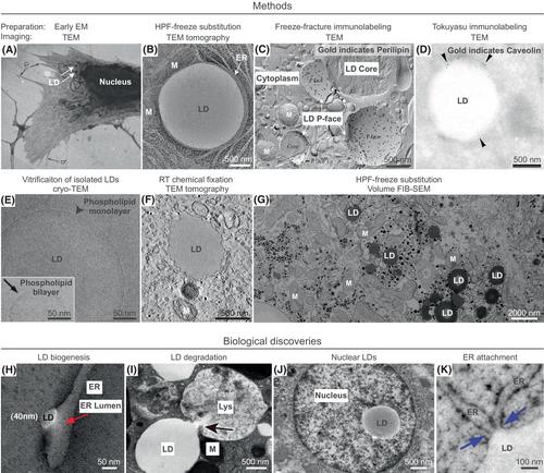
Electron microscopy (EM), in its various flavors, has significantly contributed to our understanding of lipid droplets (LD) as central organelles in cellular metabolism. For example, EM has illuminated that LDs, in contrast to all other cellular organelles, are uniquely enclosed by a single phospholipid monolayer, revealed the architecture of LD contact sites with different organelles, and provided near-atomic resolution maps of key enzymes that regulate neutral lipid biosynthesis and LD biogenesis. In this review, we first provide a brief history of pivotal findings in LD biology unveiled through the lens of an electron microscope. We describe the main EM techniques used in the context of LD research and discuss their current capabilities and limitations, thereby providing a foundation for utilizing suitable EM methodology to address LD-related questions with sufficient level of structural preservation, detail, and resolution. Finally, we highlight examples where EM has recently been and is expected to be instrumental in expanding the frontiers of LD biology.
期刊介绍:
FEBS Letters is one of the world''s leading journals in molecular biology and is renowned both for its quality of content and speed of production. Bringing together the most important developments in the molecular biosciences, FEBS Letters provides an international forum for Minireviews, Research Letters and Hypotheses that merit urgent publication.
 求助内容:
求助内容: 应助结果提醒方式:


