Ji-Seon Lee, Do-Yun Heo, Kyung-Hae Choi, Hee-Jin Kim
{"title":"Impact of the Ventricle Size on Alzheimer's Disease Progression: A Retrospective Longitudinal Study.","authors":"Ji-Seon Lee, Do-Yun Heo, Kyung-Hae Choi, Hee-Jin Kim","doi":"10.12779/dnd.2024.23.2.95","DOIUrl":null,"url":null,"abstract":"<p><strong>Background and purpose: </strong>Ventricle enlargement has been implicated in the pathophysiology of Alzheimer's disease (AD). We studied the relationship between ventricular size and cognitive function in patients with AD. We focused on the effect of the initial ventricle size on the rate of cognitive decline in patients with AD.</p><p><strong>Methods: </strong>A retrospective analysis of probable clinical AD participants with more than 2 magnetic resonance imaging images was performed. To measure ventricle size, we used visual rating scales of (1) Cardiovascular Health Study (CHS) score and (2) conventional linear measurement method.</p><p><strong>Results: </strong>Increased clinical dementia rating (CDR) was correlated with a decreased Mini-Mental Status Examination (MMSE) score, and increased medial temporal lobe atrophy (MTLA) and global ventricle size (<i>p</i><0.001, <i>p</i><0.001, <i>p</i>=0.021, respectively). There was a significant correlation between the change in cognitive function in the group (70%-100%ile) with a large initial ventricle size (<i>p</i>=0.021 for ΔCDR, <i>p</i>=0.01 for ΔMMSE), while the median ventricle size (30%-70%ile) showed correlation with other brain structural changes (MTLA, frontal atrophy [FA], and white matter) (<i>p</i>=0.036 for initial MTLA, <i>p</i>=0.034 for FA).</p><p><strong>Conclusions: </strong>In this study, the initial ventricle size may be a potential new imaging biomarker for initial cognitive function and clinical progression in AD. We found a relationship between the initial ventricle size and initial AD-related brain structural biomarkers.</p>","PeriodicalId":72779,"journal":{"name":"Dementia and neurocognitive disorders","volume":"23 2","pages":"95-106"},"PeriodicalIF":0.0000,"publicationDate":"2024-04-01","publicationTypes":"Journal Article","fieldsOfStudy":null,"isOpenAccess":false,"openAccessPdf":"https://www.ncbi.nlm.nih.gov/pmc/articles/PMC11073924/pdf/","citationCount":"0","resultStr":null,"platform":"Semanticscholar","paperid":null,"PeriodicalName":"Dementia and neurocognitive disorders","FirstCategoryId":"1085","ListUrlMain":"https://doi.org/10.12779/dnd.2024.23.2.95","RegionNum":0,"RegionCategory":null,"ArticlePicture":[],"TitleCN":null,"AbstractTextCN":null,"PMCID":null,"EPubDate":"2024/3/27 0:00:00","PubModel":"Epub","JCR":"","JCRName":"","Score":null,"Total":0}
引用次数: 0
Abstract
Background and purpose: Ventricle enlargement has been implicated in the pathophysiology of Alzheimer's disease (AD). We studied the relationship between ventricular size and cognitive function in patients with AD. We focused on the effect of the initial ventricle size on the rate of cognitive decline in patients with AD.
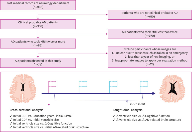
Methods: A retrospective analysis of probable clinical AD participants with more than 2 magnetic resonance imaging images was performed. To measure ventricle size, we used visual rating scales of (1) Cardiovascular Health Study (CHS) score and (2) conventional linear measurement method.
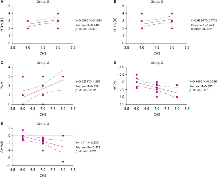
Results: Increased clinical dementia rating (CDR) was correlated with a decreased Mini-Mental Status Examination (MMSE) score, and increased medial temporal lobe atrophy (MTLA) and global ventricle size (p<0.001, p<0.001, p=0.021, respectively). There was a significant correlation between the change in cognitive function in the group (70%-100%ile) with a large initial ventricle size (p=0.021 for ΔCDR, p=0.01 for ΔMMSE), while the median ventricle size (30%-70%ile) showed correlation with other brain structural changes (MTLA, frontal atrophy [FA], and white matter) (p=0.036 for initial MTLA, p=0.034 for FA).
Conclusions: In this study, the initial ventricle size may be a potential new imaging biomarker for initial cognitive function and clinical progression in AD. We found a relationship between the initial ventricle size and initial AD-related brain structural biomarkers.
背景和目的:脑室扩大与阿尔茨海默病(AD)的病理生理学有关。我们研究了 AD 患者心室大小与认知功能之间的关系。我们重点研究了初始心室大小对阿尔茨海默病患者认知功能下降速度的影响:我们对具有 2 张以上磁共振成像图像的疑似临床 AD 患者进行了回顾性分析。测量心室大小时,我们使用了视觉评分量表(1)心血管健康研究(CHS)评分和(2)传统线性测量方法:结果:临床痴呆评分(CDR)的增加与迷你精神状态检查(MMSE)评分的降低、内侧颞叶萎缩(MTLA)的增加和总体脑室大小的增加相关(分别为 ppp=0.021)。初始脑室大小较大组(70%-100%ile)的认知功能变化与初始脑室大小有明显相关性(ΔCDR p=0.021,ΔMMSE p=0.01),而中位脑室大小(30%-70%ile)与其他脑结构变化(MTLA、额叶萎缩[FA]和白质)有相关性(初始MTLA p=0.036,FA p=0.034):在这项研究中,初始脑室大小可能是反映 AD 初始认知功能和临床进展的一种潜在的新影像生物标志物。我们发现初始脑室大小与AD相关的初始脑结构生物标志物之间存在关系。


 求助内容:
求助内容: 应助结果提醒方式:


