{"title":"Quantitative analysis of lung lesions using unenhanced chest computed tomography images","authors":"Fariba Zarei, Payam Jannatdoust, Siamak Malekpour, Mahshad Razaghi, Sabyasachi Chatterjee, Vani Varadhan Chatterjee, Amirbahador Abbasi, Rezvan Ravanfar Haghighi","doi":"10.1111/crj.13759","DOIUrl":null,"url":null,"abstract":"<div>\n \n \n <section>\n \n <h3> Introduction</h3>\n \n <p>Chest radiograph and computed tomography (CT) scans can accidentally reveal pulmonary nodules. Malignant and benign pulmonary nodules can be difficult to distinguish without specific imaging features, such as calcification, necrosis, and contrast enhancement. However, these lesions may exhibit different image texture characteristics which cannot be assessed visually. Thus, a computer-assisted quantitative method like histogram analysis (HA) of Hounsfield unit (HU) values can improve diagnostic accuracy, reducing the need for invasive biopsy.</p>\n </section>\n \n <section>\n \n <h3> Methods</h3>\n \n <p>In this exploratory control study, nonenhanced chest CT images of 20 patients with benign (10) and cancerous (10) lesion were selected retrospectively. The appearances of benign and malignant lesions were very similar in chest CT images, and only pathology report was used to discriminate them. Free hand region of interest (ROI) was inserted inside the lesion for all slices of each lesion. Mean, minimum, maximum, and standard deviations of HU values were recorded and used to make HA.</p>\n </section>\n \n <section>\n \n <h3> Results</h3>\n \n <p>HA showed that the most malignant lesions have a mean HU value between 30 and 50, a maximum HU less than 150, and a minimum HU between −30 and 20. Lesions outside these ranges were mostly benign.</p>\n </section>\n \n <section>\n \n <h3> Conclusion</h3>\n \n <p>Quantitative CT analysis may differentiate malignant from benign lesions without specific malignancy patterns on unenhanced chest CT image.</p>\n </section>\n </div>","PeriodicalId":55247,"journal":{"name":"Clinical Respiratory Journal","volume":"18 5","pages":""},"PeriodicalIF":1.9000,"publicationDate":"2024-05-07","publicationTypes":"Journal Article","fieldsOfStudy":null,"isOpenAccess":false,"openAccessPdf":"https://onlinelibrary.wiley.com/doi/epdf/10.1111/crj.13759","citationCount":"0","resultStr":null,"platform":"Semanticscholar","paperid":null,"PeriodicalName":"Clinical Respiratory Journal","FirstCategoryId":"3","ListUrlMain":"https://onlinelibrary.wiley.com/doi/10.1111/crj.13759","RegionNum":4,"RegionCategory":"医学","ArticlePicture":[],"TitleCN":null,"AbstractTextCN":null,"PMCID":null,"EPubDate":"","PubModel":"","JCR":"Q3","JCRName":"RESPIRATORY SYSTEM","Score":null,"Total":0}
引用次数: 0
Abstract
Introduction
Chest radiograph and computed tomography (CT) scans can accidentally reveal pulmonary nodules. Malignant and benign pulmonary nodules can be difficult to distinguish without specific imaging features, such as calcification, necrosis, and contrast enhancement. However, these lesions may exhibit different image texture characteristics which cannot be assessed visually. Thus, a computer-assisted quantitative method like histogram analysis (HA) of Hounsfield unit (HU) values can improve diagnostic accuracy, reducing the need for invasive biopsy.
Methods
In this exploratory control study, nonenhanced chest CT images of 20 patients with benign (10) and cancerous (10) lesion were selected retrospectively. The appearances of benign and malignant lesions were very similar in chest CT images, and only pathology report was used to discriminate them. Free hand region of interest (ROI) was inserted inside the lesion for all slices of each lesion. Mean, minimum, maximum, and standard deviations of HU values were recorded and used to make HA.
Results
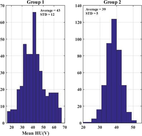
HA showed that the most malignant lesions have a mean HU value between 30 and 50, a maximum HU less than 150, and a minimum HU between −30 and 20. Lesions outside these ranges were mostly benign.
Conclusion
Quantitative CT analysis may differentiate malignant from benign lesions without specific malignancy patterns on unenhanced chest CT image.
期刊介绍:
Overview
Effective with the 2016 volume, this journal will be published in an online-only format.
Aims and Scope
The Clinical Respiratory Journal (CRJ) provides a forum for clinical research in all areas of respiratory medicine from clinical lung disease to basic research relevant to the clinic.
We publish original research, review articles, case studies, editorials and book reviews in all areas of clinical lung disease including:
Asthma
Allergy
COPD
Non-invasive ventilation
Sleep related breathing disorders
Interstitial lung diseases
Lung cancer
Clinical genetics
Rhinitis
Airway and lung infection
Epidemiology
Pediatrics
CRJ provides a fast-track service for selected Phase II and Phase III trial studies.
Keywords
Clinical Respiratory Journal, respiratory, pulmonary, medicine, clinical, lung disease,
Abstracting and Indexing Information
Academic Search (EBSCO Publishing)
Academic Search Alumni Edition (EBSCO Publishing)
Embase (Elsevier)
Health & Medical Collection (ProQuest)
Health Research Premium Collection (ProQuest)
HEED: Health Economic Evaluations Database (Wiley-Blackwell)
Hospital Premium Collection (ProQuest)
Journal Citation Reports/Science Edition (Clarivate Analytics)
MEDLINE/PubMed (NLM)
ProQuest Central (ProQuest)
Science Citation Index Expanded (Clarivate Analytics)
SCOPUS (Elsevier)
 求助内容:
求助内容: 应助结果提醒方式:


