Maria Meindl, Artem Zatcepin, Johannes Gnörich, Maximilian Scheifele, Mirlind Zaganjori, Mattes Groß, Simon Lindner, Rebecca Schaefer, Marcel Simmet, Sebastian Roemer, Sabrina Katzdobler, Johannes Levin, Günter Höglinger, Ann-Cathrin Bischof, Henryk Barthel, Osama Sabri, Peter Bartenstein, Thomas Saller, Nicolai Franzmeier, Sibylle Ziegler, Matthias Brendel
{"title":"Assessment of [<sup>18</sup>F]PI-2620 Tau-PET Quantification via Non-Invasive Automatized Image Derived Input Function.","authors":"Maria Meindl, Artem Zatcepin, Johannes Gnörich, Maximilian Scheifele, Mirlind Zaganjori, Mattes Groß, Simon Lindner, Rebecca Schaefer, Marcel Simmet, Sebastian Roemer, Sabrina Katzdobler, Johannes Levin, Günter Höglinger, Ann-Cathrin Bischof, Henryk Barthel, Osama Sabri, Peter Bartenstein, Thomas Saller, Nicolai Franzmeier, Sibylle Ziegler, Matthias Brendel","doi":"10.1007/s00259-024-06741-7","DOIUrl":null,"url":null,"abstract":"<p><strong>Purpose: </strong>[<sup>18</sup>F]PI-2620 positron emission tomography (PET) detects misfolded tau in progressive supranuclear palsy (PSP) and Alzheimer's disease (AD). We questioned the feasibility and value of absolute [<sup>18</sup>F]PI-2620 PET quantification for assessing tau by regional distribution volumes (V<sub>T</sub>). Here, arterial input functions (AIF) represent the gold standard, but cannot be applied in routine clinical practice, whereas image-derived input functions (IDIF) represent a non-invasive alternative. We aimed to validate IDIF against AIF and we evaluated the potential to discriminate patients with PSP and AD from healthy controls by non-invasive quantification of [<sup>18</sup>F] PET.</p><p><strong>Methods: </strong>In the first part of the study, we validated AIF derived from radial artery whole blood against IDIF by investigating 20 subjects (ten controls and ten patients). IDIF were generated by manual extraction of the carotid artery using the average and the five highest (max5) voxel intensity values and by automated extraction of the carotid artery using the average and the maximum voxel intensity value. In the second part of the study, IDIF quantification using the IDIF with the closest match to the AIF was transferred to group comparison of a large independent cohort of 40 subjects (15 healthy controls, 15 PSP patients and 10 AD patients). We compared V<sub>T</sub> and V<sub>T</sub> ratios, both calculated by Logan plots, with distribution volume (DV) ratios using simplified reference tissue modelling and standardized uptake value (SUV) ratios.</p><p><strong>Results: </strong>AIF and IDIF showed highly correlated input curves for all applied IDIF extraction methods (0.78 < r < 0.83, all p < 0.0001; area under the curves (AUC): 0.73 < r ≤ 0.82, all p ≤ 0.0003). Regarding the V<sub>T</sub> values, correlations were mainly found between those generated by the AIF and by the IDIF methods using the maximum voxel intensity values. Lowest relative differences (RD) were observed by applying the manual method using the five highest voxel intensity values (max5) (AIF vs. IDIF manual, avg: RD = -82%; AIF vs. IDIF automated, avg: RD = -86%; AIF vs. IDIF manual, max5: RD = -6%; AIF vs. IDIF automated, max: RD = -26%). Regional V<sub>T</sub> values revealed considerable variance at group level, which was strongly reduced upon scaling by the inferior cerebellum. The resulting V<sub>T</sub> ratio values were adequate to detect group differences between patients with PSP or AD and healthy controls (HC) (PSP target region (globus pallidus): HC vs. PSP vs. AD: 1.18 vs. 1.32 vs. 1.16; AD target region (Braak region I): HC vs. PSP vs. AD: 1.00 vs. 1.00 vs. 1.22). V<sub>T</sub> ratios and DV ratios outperformed SUV ratios and V<sub>T</sub> in detecting differences between PSP and healthy controls, whereas all quantification approaches performed similarly in comparing AD and healthy controls.</p><p><strong>Conclusion: </strong>Blood-free IDIF is a promising approach for quantification of [<sup>18</sup>F]PI-2620 PET, serving as correlating surrogate for invasive continuous arterial blood sampling. Regional [<sup>18</sup>F]PI-2620 V<sub>T</sub> show large variance, in contrast to regional [<sup>18</sup>F]PI-2620 V<sub>T</sub> ratios scaled with the inferior cerebellum, which are appropriate for discriminating PSP, AD and healthy controls. DV ratios obtained by simplified reference tissue modeling are similarly suitable for this purpose.</p>","PeriodicalId":11909,"journal":{"name":"European Journal of Nuclear Medicine and Molecular Imaging","volume":null,"pages":null},"PeriodicalIF":8.6000,"publicationDate":"2024-09-01","publicationTypes":"Journal Article","fieldsOfStudy":null,"isOpenAccess":false,"openAccessPdf":"https://www.ncbi.nlm.nih.gov/pmc/articles/PMC11368995/pdf/","citationCount":"0","resultStr":null,"platform":"Semanticscholar","paperid":null,"PeriodicalName":"European Journal of Nuclear Medicine and Molecular Imaging","FirstCategoryId":"3","ListUrlMain":"https://doi.org/10.1007/s00259-024-06741-7","RegionNum":1,"RegionCategory":"医学","ArticlePicture":[],"TitleCN":null,"AbstractTextCN":null,"PMCID":null,"EPubDate":"2024/5/8 0:00:00","PubModel":"Epub","JCR":"Q1","JCRName":"RADIOLOGY, NUCLEAR MEDICINE & MEDICAL IMAGING","Score":null,"Total":0}
引用次数: 0
Abstract
Purpose: [18F]PI-2620 positron emission tomography (PET) detects misfolded tau in progressive supranuclear palsy (PSP) and Alzheimer's disease (AD). We questioned the feasibility and value of absolute [18F]PI-2620 PET quantification for assessing tau by regional distribution volumes (VT). Here, arterial input functions (AIF) represent the gold standard, but cannot be applied in routine clinical practice, whereas image-derived input functions (IDIF) represent a non-invasive alternative. We aimed to validate IDIF against AIF and we evaluated the potential to discriminate patients with PSP and AD from healthy controls by non-invasive quantification of [18F] PET.
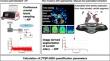
Methods: In the first part of the study, we validated AIF derived from radial artery whole blood against IDIF by investigating 20 subjects (ten controls and ten patients). IDIF were generated by manual extraction of the carotid artery using the average and the five highest (max5) voxel intensity values and by automated extraction of the carotid artery using the average and the maximum voxel intensity value. In the second part of the study, IDIF quantification using the IDIF with the closest match to the AIF was transferred to group comparison of a large independent cohort of 40 subjects (15 healthy controls, 15 PSP patients and 10 AD patients). We compared VT and VT ratios, both calculated by Logan plots, with distribution volume (DV) ratios using simplified reference tissue modelling and standardized uptake value (SUV) ratios.
Results: AIF and IDIF showed highly correlated input curves for all applied IDIF extraction methods (0.78 < r < 0.83, all p < 0.0001; area under the curves (AUC): 0.73 < r ≤ 0.82, all p ≤ 0.0003). Regarding the VT values, correlations were mainly found between those generated by the AIF and by the IDIF methods using the maximum voxel intensity values. Lowest relative differences (RD) were observed by applying the manual method using the five highest voxel intensity values (max5) (AIF vs. IDIF manual, avg: RD = -82%; AIF vs. IDIF automated, avg: RD = -86%; AIF vs. IDIF manual, max5: RD = -6%; AIF vs. IDIF automated, max: RD = -26%). Regional VT values revealed considerable variance at group level, which was strongly reduced upon scaling by the inferior cerebellum. The resulting VT ratio values were adequate to detect group differences between patients with PSP or AD and healthy controls (HC) (PSP target region (globus pallidus): HC vs. PSP vs. AD: 1.18 vs. 1.32 vs. 1.16; AD target region (Braak region I): HC vs. PSP vs. AD: 1.00 vs. 1.00 vs. 1.22). VT ratios and DV ratios outperformed SUV ratios and VT in detecting differences between PSP and healthy controls, whereas all quantification approaches performed similarly in comparing AD and healthy controls.
Conclusion: Blood-free IDIF is a promising approach for quantification of [18F]PI-2620 PET, serving as correlating surrogate for invasive continuous arterial blood sampling. Regional [18F]PI-2620 VT show large variance, in contrast to regional [18F]PI-2620 VT ratios scaled with the inferior cerebellum, which are appropriate for discriminating PSP, AD and healthy controls. DV ratios obtained by simplified reference tissue modeling are similarly suitable for this purpose.
期刊介绍:
The European Journal of Nuclear Medicine and Molecular Imaging serves as a platform for the exchange of clinical and scientific information within nuclear medicine and related professions. It welcomes international submissions from professionals involved in the functional, metabolic, and molecular investigation of diseases. The journal's coverage spans physics, dosimetry, radiation biology, radiochemistry, and pharmacy, providing high-quality peer review by experts in the field. Known for highly cited and downloaded articles, it ensures global visibility for research work and is part of the EJNMMI journal family.

 求助内容:
求助内容: 应助结果提醒方式:


