Mohammad Maskani, Samaneh Abbasi, Hamidreza Etemad-Rezaee, Hamid Abdolahi, Amir Zamanpour, Alireza Montazerabadi
{"title":"Grading of Gliomas by Contrast-Enhanced CT Radiomics Features.","authors":"Mohammad Maskani, Samaneh Abbasi, Hamidreza Etemad-Rezaee, Hamid Abdolahi, Amir Zamanpour, Alireza Montazerabadi","doi":"10.31661/jbpe.v0i0.2306-1628","DOIUrl":null,"url":null,"abstract":"<p><strong>Background: </strong>Gliomas, as Central Nervous System (CNS) tumors, are greatly common with 80% of malignancy. Treatment methods for gliomas, such as surgery, radiation therapy, and chemotherapy depend on the grade, size, location, and the patient's age.</p><p><strong>Objective: </strong>This study aimed to quantify glioma based on the radiomics analysis and classify its grade into High-grade Glioma (HGG) or Low-grade Glioma (LGG) by various machine-learning methods using contrast-enhanced brain Computerized Tomography (CT) scans.</p><p><strong>Material and methods: </strong>This retrospective study involved acquiring and segmenting data, selecting and extracting features, classifying, analyzing, and evaluating classifiers. The study included a total of 62 patients (31 with LGG and 31 with HGG). The tumors were segmented by an experienced CT-scan technologist with 3D slicer software. A total of 14 shape features, 18 histogram-based features, and 75 texture-based features were computed. The Area Under the Curve (AUC) and Receiver Operating Characteristic Curve (ROC) were used to evaluate and compare classification models.</p><p><strong>Results: </strong>A total of 13 out of 107 features were selected to differentiate between LGGs and HGGs and to perform various classifier algorithms with different cross-validations. The best classifier algorithm was linear-discriminant with 93.5% accuracy, 96.77% sensitivity, 90.3% specificity, and 0.98% AUC in the differentiation of LGGs and HGGs.</p><p><strong>Conclusion: </strong>The proposed method can identify LGG and HGG with 93.5% accuracy, 96.77% sensitivity, 90.3% specificity, and 0.98% AUC, leading to the best treatment for glioma patients by using CT scans based on radiomics analysis.</p>","PeriodicalId":38035,"journal":{"name":"Journal of Biomedical Physics and Engineering","volume":"14 2","pages":"151-158"},"PeriodicalIF":0.0000,"publicationDate":"2024-04-01","publicationTypes":"Journal Article","fieldsOfStudy":null,"isOpenAccess":false,"openAccessPdf":"https://www.ncbi.nlm.nih.gov/pmc/articles/PMC11016825/pdf/","citationCount":"0","resultStr":null,"platform":"Semanticscholar","paperid":null,"PeriodicalName":"Journal of Biomedical Physics and Engineering","FirstCategoryId":"1085","ListUrlMain":"https://doi.org/10.31661/jbpe.v0i0.2306-1628","RegionNum":0,"RegionCategory":null,"ArticlePicture":[],"TitleCN":null,"AbstractTextCN":null,"PMCID":null,"EPubDate":"","PubModel":"","JCR":"Q3","JCRName":"Medicine","Score":null,"Total":0}
引用次数: 0
Abstract
Background: Gliomas, as Central Nervous System (CNS) tumors, are greatly common with 80% of malignancy. Treatment methods for gliomas, such as surgery, radiation therapy, and chemotherapy depend on the grade, size, location, and the patient's age.
Objective: This study aimed to quantify glioma based on the radiomics analysis and classify its grade into High-grade Glioma (HGG) or Low-grade Glioma (LGG) by various machine-learning methods using contrast-enhanced brain Computerized Tomography (CT) scans.
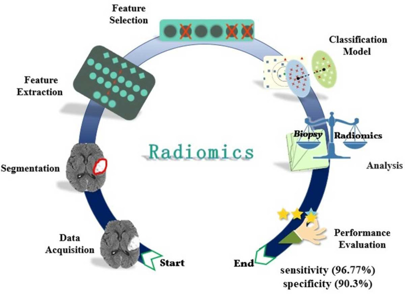
Material and methods: This retrospective study involved acquiring and segmenting data, selecting and extracting features, classifying, analyzing, and evaluating classifiers. The study included a total of 62 patients (31 with LGG and 31 with HGG). The tumors were segmented by an experienced CT-scan technologist with 3D slicer software. A total of 14 shape features, 18 histogram-based features, and 75 texture-based features were computed. The Area Under the Curve (AUC) and Receiver Operating Characteristic Curve (ROC) were used to evaluate and compare classification models.
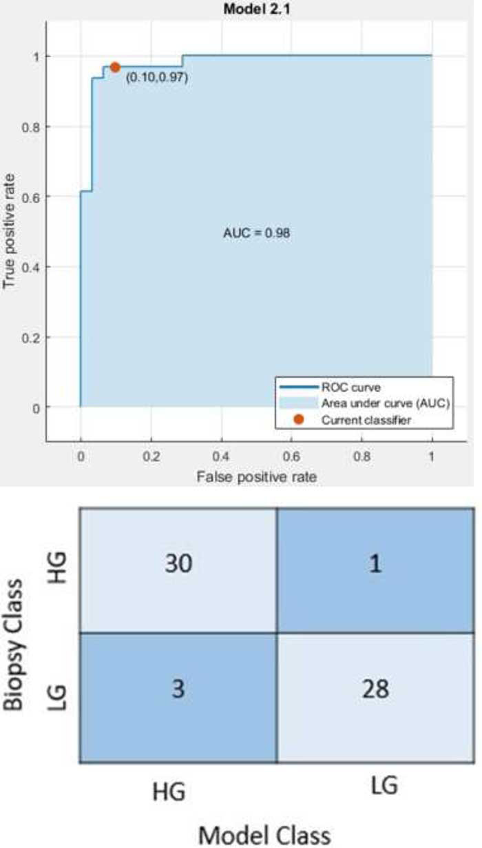
Results: A total of 13 out of 107 features were selected to differentiate between LGGs and HGGs and to perform various classifier algorithms with different cross-validations. The best classifier algorithm was linear-discriminant with 93.5% accuracy, 96.77% sensitivity, 90.3% specificity, and 0.98% AUC in the differentiation of LGGs and HGGs.
Conclusion: The proposed method can identify LGG and HGG with 93.5% accuracy, 96.77% sensitivity, 90.3% specificity, and 0.98% AUC, leading to the best treatment for glioma patients by using CT scans based on radiomics analysis.
期刊介绍:
The Journal of Biomedical Physics and Engineering (JBPE) is a bimonthly peer-reviewed English-language journal that publishes high-quality basic sciences and clinical research (experimental or theoretical) broadly concerned with the relationship of physics to medicine and engineering.

 求助内容:
求助内容: 应助结果提醒方式:


