Gaurav Ambwani, Zhongjie Shi, Kehuan Luo, Jeong-Won Jeong, Sidhartha Tan
{"title":"Distinguishing Laterality in Brain Injury in Rabbit Fetal Magnetic Resonance Imaging Using Novel Volume Rendering Techniques.","authors":"Gaurav Ambwani, Zhongjie Shi, Kehuan Luo, Jeong-Won Jeong, Sidhartha Tan","doi":"10.1159/000539212","DOIUrl":null,"url":null,"abstract":"<p><strong>Introduction: </strong>Our laboratory has been exploring the MRI detection of fetal brain injury, which previously provided a prognostic biomarker for newborn hypertonia in an animal model of cerebral palsy (CP). The biomarker relies on distinct patterns of diffusion-weighted imaging-defined apparent diffusion coefficient (ADC) in fetal brains during uterine hypoxia-ischemia (H-I). Despite the challenges posed by small brains and tissue acquisition, our objective was to differentiate between left and right brain ADC changes.</p><p><strong>Methods: </strong>A novel aspect involved utilizing three-dimensional rendering techniques to refine ADC measurements within spheroids encompassing fetal brain tissue. 25-day gestation age of rabbit fetuses underwent global hypoxia due to maternal uterine ischemia.</p><p><strong>Results: </strong>Successful differentiation of left and right brain regions was achieved in 28% of the fetal brains. Ordinal analysis revealed predominantly higher ADC on the left side compared to the right at baseline and across the entire time series. During H-I and reperfusion-reoxygenation, the right side exhibited a favored percentage change. Among these fetal brains, 73% exhibited the ADC pattern predictive of hypertonia. No significant differences between left and right sides were observed in patterns predicting hypertonia, except for one timepoint during H-I. This study also highlights a balance between left-sided and right-sided alterations within the population.</p><p><strong>Conclusion: </strong>This study emphasizes the importance of investigating laterality and asymmetric hemispheric lesions for early diagnosis of brain injury, leading to CP. The technological limitations in obtaining a clear picture of the entire fetal brain for every fetus mirror the challenges encountered in human studies.</p><p><strong>Introduction: </strong>Our laboratory has been exploring the MRI detection of fetal brain injury, which previously provided a prognostic biomarker for newborn hypertonia in an animal model of cerebral palsy (CP). The biomarker relies on distinct patterns of diffusion-weighted imaging-defined apparent diffusion coefficient (ADC) in fetal brains during uterine hypoxia-ischemia (H-I). Despite the challenges posed by small brains and tissue acquisition, our objective was to differentiate between left and right brain ADC changes.</p><p><strong>Methods: </strong>A novel aspect involved utilizing three-dimensional rendering techniques to refine ADC measurements within spheroids encompassing fetal brain tissue. 25-day gestation age of rabbit fetuses underwent global hypoxia due to maternal uterine ischemia.</p><p><strong>Results: </strong>Successful differentiation of left and right brain regions was achieved in 28% of the fetal brains. Ordinal analysis revealed predominantly higher ADC on the left side compared to the right at baseline and across the entire time series. During H-I and reperfusion-reoxygenation, the right side exhibited a favored percentage change. Among these fetal brains, 73% exhibited the ADC pattern predictive of hypertonia. No significant differences between left and right sides were observed in patterns predicting hypertonia, except for one timepoint during H-I. This study also highlights a balance between left-sided and right-sided alterations within the population.</p><p><strong>Conclusion: </strong>This study emphasizes the importance of investigating laterality and asymmetric hemispheric lesions for early diagnosis of brain injury, leading to CP. The technological limitations in obtaining a clear picture of the entire fetal brain for every fetus mirror the challenges encountered in human studies.</p>","PeriodicalId":50585,"journal":{"name":"Developmental Neuroscience","volume":" ","pages":"55-67"},"PeriodicalIF":2.0000,"publicationDate":"2025-01-01","publicationTypes":"Journal Article","fieldsOfStudy":null,"isOpenAccess":false,"openAccessPdf":"https://www.ncbi.nlm.nih.gov/pmc/articles/PMC11538374/pdf/","citationCount":"0","resultStr":null,"platform":"Semanticscholar","paperid":null,"PeriodicalName":"Developmental Neuroscience","FirstCategoryId":"3","ListUrlMain":"https://doi.org/10.1159/000539212","RegionNum":4,"RegionCategory":"医学","ArticlePicture":[],"TitleCN":null,"AbstractTextCN":null,"PMCID":null,"EPubDate":"2024/5/6 0:00:00","PubModel":"Epub","JCR":"Q2","JCRName":"DEVELOPMENTAL BIOLOGY","Score":null,"Total":0}
引用次数: 0
Abstract
Introduction: Our laboratory has been exploring the MRI detection of fetal brain injury, which previously provided a prognostic biomarker for newborn hypertonia in an animal model of cerebral palsy (CP). The biomarker relies on distinct patterns of diffusion-weighted imaging-defined apparent diffusion coefficient (ADC) in fetal brains during uterine hypoxia-ischemia (H-I). Despite the challenges posed by small brains and tissue acquisition, our objective was to differentiate between left and right brain ADC changes.
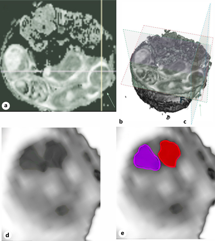
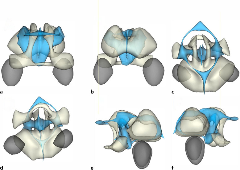
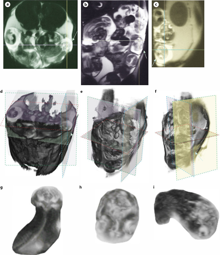
Methods: A novel aspect involved utilizing three-dimensional rendering techniques to refine ADC measurements within spheroids encompassing fetal brain tissue. 25-day gestation age of rabbit fetuses underwent global hypoxia due to maternal uterine ischemia.
Results: Successful differentiation of left and right brain regions was achieved in 28% of the fetal brains. Ordinal analysis revealed predominantly higher ADC on the left side compared to the right at baseline and across the entire time series. During H-I and reperfusion-reoxygenation, the right side exhibited a favored percentage change. Among these fetal brains, 73% exhibited the ADC pattern predictive of hypertonia. No significant differences between left and right sides were observed in patterns predicting hypertonia, except for one timepoint during H-I. This study also highlights a balance between left-sided and right-sided alterations within the population.
Conclusion: This study emphasizes the importance of investigating laterality and asymmetric hemispheric lesions for early diagnosis of brain injury, leading to CP. The technological limitations in obtaining a clear picture of the entire fetal brain for every fetus mirror the challenges encountered in human studies.
Introduction: Our laboratory has been exploring the MRI detection of fetal brain injury, which previously provided a prognostic biomarker for newborn hypertonia in an animal model of cerebral palsy (CP). The biomarker relies on distinct patterns of diffusion-weighted imaging-defined apparent diffusion coefficient (ADC) in fetal brains during uterine hypoxia-ischemia (H-I). Despite the challenges posed by small brains and tissue acquisition, our objective was to differentiate between left and right brain ADC changes.
Methods: A novel aspect involved utilizing three-dimensional rendering techniques to refine ADC measurements within spheroids encompassing fetal brain tissue. 25-day gestation age of rabbit fetuses underwent global hypoxia due to maternal uterine ischemia.
Results: Successful differentiation of left and right brain regions was achieved in 28% of the fetal brains. Ordinal analysis revealed predominantly higher ADC on the left side compared to the right at baseline and across the entire time series. During H-I and reperfusion-reoxygenation, the right side exhibited a favored percentage change. Among these fetal brains, 73% exhibited the ADC pattern predictive of hypertonia. No significant differences between left and right sides were observed in patterns predicting hypertonia, except for one timepoint during H-I. This study also highlights a balance between left-sided and right-sided alterations within the population.
Conclusion: This study emphasizes the importance of investigating laterality and asymmetric hemispheric lesions for early diagnosis of brain injury, leading to CP. The technological limitations in obtaining a clear picture of the entire fetal brain for every fetus mirror the challenges encountered in human studies.
期刊介绍:
''Developmental Neuroscience'' is a multidisciplinary journal publishing papers covering all stages of invertebrate, vertebrate and human brain development. Emphasis is placed on publishing fundamental as well as translational studies that contribute to our understanding of mechanisms of normal development as well as genetic and environmental causes of abnormal brain development. The journal thus provides valuable information for both physicians and biologists. To meet the rapidly expanding information needs of its readers, the journal combines original papers that report on progress and advances in developmental neuroscience with concise mini-reviews that provide a timely overview of key topics, new insights and ongoing controversies. The editorial standards of ''Developmental Neuroscience'' are high. We are committed to publishing only high quality, complete papers that make significant contributions to the field.
 求助内容:
求助内容: 应助结果提醒方式:


