The histological spectrum and immunoprofile of head and neck NUT carcinoma: A multicentre series of 30 cases
Abstract
Background and aim
Head and neck nuclear protein of testis carcinoma (HN-NUT) is a rare form of carcinoma diagnosed by NUT immunohistochemistry positivity and/or NUTM1 translocation. Although the prototype of HN-NUT is a primitive undifferentiated round cell tumour (URC) with immunopositivity for squamous markers, it is our observation that it may assume variant histology or immunoprofile.
Methods
We conducted a detailed clinicopathological review of a large retrospective cohort of 30 HN-NUT, aiming to expand its histological and immunohistochemical spectrum.
Results
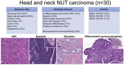
The median age of patients with HN-NUT was 39 years (range = 17–86). It affected the sinonasal tract (43%), major salivary glands (20%), thyroid (13%), oral cavity (7%), larynx (7%), neck (7%) and nasopharynx (3%). Although most cases of HN-NUT (63%) contained a component of primitive URC tumour, 53% showed other histological features and 37% lacked a URC component altogether. Variant histological features included basaloid (33%), differentiated squamous/squamoid (37%), clear cell changes (13%), glandular differentiation (7%) and papillary architecture (10%), which could co-exist. While most HN-NUT were positive for keratins, p63 and p40, occasional cases (5–9%) were entirely negative. Immunopositivity for neuroendocrine markers and thyroid transcription factor-1 was observed in 33 and 36% of cases, respectively. The outcome of HN-NUT was dismal, with a 3-year disease specific survival of 38%.
Conclusions
HN-NUT can affect individuals across a wide age range and arise from various head and neck sites. It exhibits a diverse spectrum of histological features and may be positive for neuroendocrine markers, potentially leading to underdiagnosis. A low threshold to perform NUT-specific tests is necessary to accurately diagnose HN-NUT.
| 公司名称 | 产品信息 | 采购帮参考价格 |
|---|
 求助内容:
求助内容: 应助结果提醒方式:


