{"title":"An atypical case involving real, ghost, and pseudo-ghost images on a panoramic radiograph.","authors":"Jong-Won Kim, Yo-Seob Seo","doi":"10.5624/isd.20230256","DOIUrl":null,"url":null,"abstract":"<p><strong>Purpose: </strong>This report presents a unique case featuring real, ghost, and pseudo-ghost images on the panoramic radiograph of a patient wearing earrings. It also explains the formation of these images in an easy-to-understand manner.</p><p><strong>Material and methods: </strong>One real image and two ghost images appeared on each side of a panoramic radiograph of a patient wearing earrings on both sides. Of the two ghost images on each side, one was considered a typical ghost image and the other was considered a ghost-like real image (pseudo-ghost image). The formation zones of the real, double, and ghost images were examined based on the path and angles of the X-ray beam from the Planmeca ProMax. To simulate the pseudo-ghost and typical ghost images on panoramic radiography, a radiopaque marker was affixed to the right mandibular condyle of a dry mandible, and the position of the mandible was adjusted accordingly.</p><p><strong>Results: </strong>The center of rotation of the Planmeca ProMax extended beyond the jaw area, and the area of double image formation also reached beyond the jaw. The radiopaque-marked mandibular condyle, situated in the outwardly extending area of double image formation, exhibited triple images consisting of real, double (pseudo-ghost), and ghost images. These findings helped to explain the image formation associated with the patient's earrings observed in the panoramic radiograph.</p><p><strong>Conclusion: </strong>Dentists must understand the characteristics and principles of the panoramic equipment they use and apply this understanding to taking and interpreting panoramic radiographs.</p>","PeriodicalId":51714,"journal":{"name":"Imaging Science in Dentistry","volume":"54 1","pages":"57-62"},"PeriodicalIF":2.1000,"publicationDate":"2024-03-01","publicationTypes":"Journal Article","fieldsOfStudy":null,"isOpenAccess":false,"openAccessPdf":"https://www.ncbi.nlm.nih.gov/pmc/articles/PMC10985519/pdf/","citationCount":"0","resultStr":null,"platform":"Semanticscholar","paperid":null,"PeriodicalName":"Imaging Science in Dentistry","FirstCategoryId":"1085","ListUrlMain":"https://doi.org/10.5624/isd.20230256","RegionNum":0,"RegionCategory":null,"ArticlePicture":[],"TitleCN":null,"AbstractTextCN":null,"PMCID":null,"EPubDate":"2024/2/6 0:00:00","PubModel":"Epub","JCR":"Q3","JCRName":"DENTISTRY, ORAL SURGERY & MEDICINE","Score":null,"Total":0}
引用次数: 0
Abstract
Purpose: This report presents a unique case featuring real, ghost, and pseudo-ghost images on the panoramic radiograph of a patient wearing earrings. It also explains the formation of these images in an easy-to-understand manner.
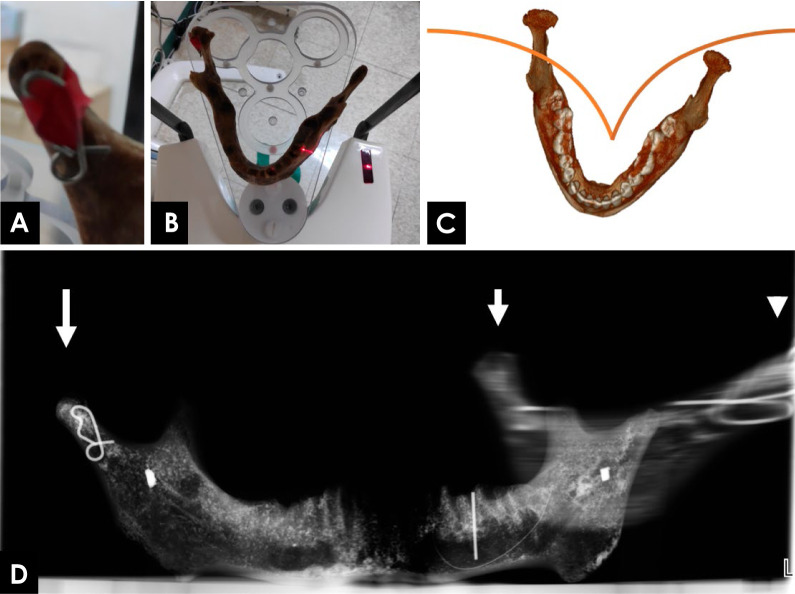
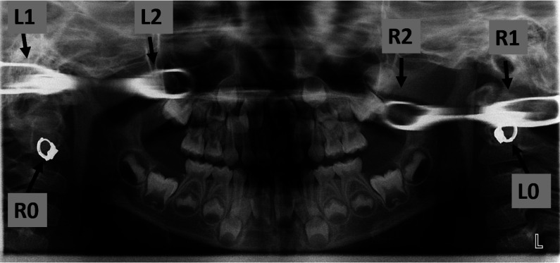
Material and methods: One real image and two ghost images appeared on each side of a panoramic radiograph of a patient wearing earrings on both sides. Of the two ghost images on each side, one was considered a typical ghost image and the other was considered a ghost-like real image (pseudo-ghost image). The formation zones of the real, double, and ghost images were examined based on the path and angles of the X-ray beam from the Planmeca ProMax. To simulate the pseudo-ghost and typical ghost images on panoramic radiography, a radiopaque marker was affixed to the right mandibular condyle of a dry mandible, and the position of the mandible was adjusted accordingly.
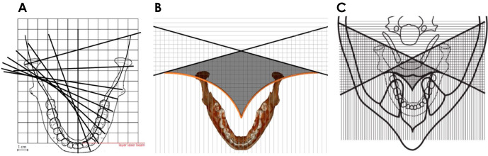
Results: The center of rotation of the Planmeca ProMax extended beyond the jaw area, and the area of double image formation also reached beyond the jaw. The radiopaque-marked mandibular condyle, situated in the outwardly extending area of double image formation, exhibited triple images consisting of real, double (pseudo-ghost), and ghost images. These findings helped to explain the image formation associated with the patient's earrings observed in the panoramic radiograph.
Conclusion: Dentists must understand the characteristics and principles of the panoramic equipment they use and apply this understanding to taking and interpreting panoramic radiographs.
目的:本报告介绍了一个独特的病例,该病例的特点是佩戴耳环的患者的全景 X 光片上出现了真实、鬼魂和假鬼魂图像。材料和方法:材料:一名佩戴耳环的患者的两侧全景 X 光片上各出现了一个真实图像和两个幽灵图像。在每侧的两个鬼影中,一个被认为是典型的鬼影,另一个被认为是类似鬼影的真影(伪鬼影)。根据 Planmeca ProMax 的 X 射线光束的路径和角度,检查了真实图像、双重图像和鬼影图像的形成区域。为了在全景放射摄影中模拟伪鬼影和典型鬼影,在干燥下颌骨的右下颌骨髁突上贴了一个不透射线的标记,并相应地调整了下颌骨的位置:结果:Planmeca ProMax 的旋转中心超出了下颌区域,双图像形成区域也超出了下颌。位于双图像形成区域向外延伸的下颌骨髁状突放射标记显示出由真实图像、双图像(伪鬼图像)和鬼图像组成的三重图像。这些发现有助于解释全景X光片上观察到的与患者耳环相关的影像形成:牙医必须了解他们所使用的全景设备的特点和原理,并在拍摄和解释全景 X 光片时应用这种理解。

 求助内容:
求助内容: 应助结果提醒方式:


