{"title":"Acute muscle loss assessed using panoramic ultrasound in critically ill adults: a prospective observational study.","authors":"Daisuke Ikechi, Hidehiko Nakano, Nobuto Nakanishi, Takahiro Fujita, Naho Watanabe, Yasuaki Koyama, Hideki Hashimoto, Kensuke Nakamura","doi":"10.1007/s10396-024-01412-4","DOIUrl":null,"url":null,"abstract":"<p><strong>Purpose: </strong>Panoramic ultrasound is one of the recently introduced ultrasound evaluation techniques. We herein examined the relationship between the cross-sectional area of the rectus femoris muscle on panoramic ultrasound and its volume based on the gold standard computed tomography (CT) evaluation.</p><p><strong>Methods: </strong>This was a single-center prospective observational study. A panoramic ultrasound assessment of the cross-sectional area of the rectus femoris muscle and a simple CT evaluation of its volume were performed on days 1 and 7 of hospitalization. Physical functions were assessed at discharge.</p><p><strong>Results: </strong>Twenty patients were examined. The rate of change in the cross-sectional area of the rectus femoris muscle on panoramic ultrasound correlated with that in its volume on CT (correlation coefficient 0.59, p = 0.0061). In addition, a correlation was observed between the absolute value for the rectus femoris muscle cross-sectional area on panoramic ultrasound and physical functions at discharge. Rectus femoris muscle distances did not correlate with either.</p><p><strong>Conclusion: </strong>In the acute phase of critical illness, the cross-sectional area of the rectus femoris muscle on panoramic images correlated with its volume on CT and, thus, it is a valid method for assessing muscle mass.</p>","PeriodicalId":50130,"journal":{"name":"Journal of Medical Ultrasonics","volume":null,"pages":null},"PeriodicalIF":1.9000,"publicationDate":"2024-04-01","publicationTypes":"Journal Article","fieldsOfStudy":null,"isOpenAccess":false,"openAccessPdf":"","citationCount":"0","resultStr":null,"platform":"Semanticscholar","paperid":null,"PeriodicalName":"Journal of Medical Ultrasonics","FirstCategoryId":"3","ListUrlMain":"https://doi.org/10.1007/s10396-024-01412-4","RegionNum":4,"RegionCategory":"医学","ArticlePicture":[],"TitleCN":null,"AbstractTextCN":null,"PMCID":null,"EPubDate":"2024/5/3 0:00:00","PubModel":"Epub","JCR":"Q3","JCRName":"RADIOLOGY, NUCLEAR MEDICINE & MEDICAL IMAGING","Score":null,"Total":0}
引用次数: 0
Abstract
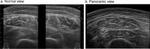
Purpose: Panoramic ultrasound is one of the recently introduced ultrasound evaluation techniques. We herein examined the relationship between the cross-sectional area of the rectus femoris muscle on panoramic ultrasound and its volume based on the gold standard computed tomography (CT) evaluation.
Methods: This was a single-center prospective observational study. A panoramic ultrasound assessment of the cross-sectional area of the rectus femoris muscle and a simple CT evaluation of its volume were performed on days 1 and 7 of hospitalization. Physical functions were assessed at discharge.
Results: Twenty patients were examined. The rate of change in the cross-sectional area of the rectus femoris muscle on panoramic ultrasound correlated with that in its volume on CT (correlation coefficient 0.59, p = 0.0061). In addition, a correlation was observed between the absolute value for the rectus femoris muscle cross-sectional area on panoramic ultrasound and physical functions at discharge. Rectus femoris muscle distances did not correlate with either.
Conclusion: In the acute phase of critical illness, the cross-sectional area of the rectus femoris muscle on panoramic images correlated with its volume on CT and, thus, it is a valid method for assessing muscle mass.
期刊介绍:
The Journal of Medical Ultrasonics is the official journal of the Japan Society of Ultrasonics in Medicine. The main purpose of the journal is to provide forum for the publication of papers documenting recent advances and new developments in the entire field of ultrasound in medicine and biology, encompassing both the medical and the engineering aspects of the science.The journal welcomes original articles, review articles, images, and letters to the editor.The journal also provides state-of-the-art information such as announcements from the boards and the committees of the society.
 求助内容:
求助内容: 应助结果提醒方式:


