Rashmitha, K. N. Manjunath, Anjali Kulkarni, Vamshikrishna Kulkarni
{"title":"Segmentation and Volumetric Analysis of Heart from Cardiac CT Images","authors":"Rashmitha, K. N. Manjunath, Anjali Kulkarni, Vamshikrishna Kulkarni","doi":"10.1007/s13239-024-00715-4","DOIUrl":null,"url":null,"abstract":"<h3 data-test=\"abstract-sub-heading\">Purpose</h3><p>Cardiac CT is a valuable diagnostic tool in evaluating cardiovascular diseases. Accurate segmentation of the heart and its structures from cardiac CT and MRI images is essential for diagnosing functional abnormalities, treatment plans and cardiovascular diseases management. Accurate segmentation and quantitative assessments are still a challenge. Manual delineation of the heart from the scan images is labour-intensive, time-consuming, and error prone as it depends on the radiologist's experience. Thus, automated techniques are highly desirable as they can significantly improve the efficiency and accuracy of image analysis.</p><h3 data-test=\"abstract-sub-heading\">Method</h3><p>This work addresses the above problems. A new, image-driven, fast, and fully automatic segmentation method was developed to segment the heart from CT images using a processing pipeline of adaptive median filter, multi-level thresholding, active contours, mathematical morphology, and the knowledge of human anatomy to delineate the regions of interest.</p><h3 data-test=\"abstract-sub-heading\">Results</h3><p>The algorithm proposed is simple to implement and validate and requires no human intervention. The method is tested on the 'Image CHD' DICOM images (multi-centre, clinically approved single-phase de-identified images), and the results obtained were validated against the ground truths provided with the dataset. The results show an average Dice score, Jaccard score, and Hausdorff distance of <i>0.866, 0.776</i>, and <i>33.29 mm</i>, respectively, for the segmentation of the heart's chambers, aorta, and blood vessels. The results and the ground truths were compared using Bland-Altmon plots.</p><h3 data-test=\"abstract-sub-heading\">Conclusion</h3><p>The heart was correctly segmented from the CT images using the proposed method. Further this segmentation technique can be used to develop AI based solutions for segmentation.</p>","PeriodicalId":54322,"journal":{"name":"Cardiovascular Engineering and Technology","volume":"16 1","pages":""},"PeriodicalIF":1.8000,"publicationDate":"2024-04-30","publicationTypes":"Journal Article","fieldsOfStudy":null,"isOpenAccess":false,"openAccessPdf":"","citationCount":"0","resultStr":null,"platform":"Semanticscholar","paperid":null,"PeriodicalName":"Cardiovascular Engineering and Technology","FirstCategoryId":"5","ListUrlMain":"https://doi.org/10.1007/s13239-024-00715-4","RegionNum":4,"RegionCategory":"医学","ArticlePicture":[],"TitleCN":null,"AbstractTextCN":null,"PMCID":null,"EPubDate":"","PubModel":"","JCR":"Q3","JCRName":"CARDIAC & CARDIOVASCULAR SYSTEMS","Score":null,"Total":0}
引用次数: 0
Abstract
Purpose
Cardiac CT is a valuable diagnostic tool in evaluating cardiovascular diseases. Accurate segmentation of the heart and its structures from cardiac CT and MRI images is essential for diagnosing functional abnormalities, treatment plans and cardiovascular diseases management. Accurate segmentation and quantitative assessments are still a challenge. Manual delineation of the heart from the scan images is labour-intensive, time-consuming, and error prone as it depends on the radiologist's experience. Thus, automated techniques are highly desirable as they can significantly improve the efficiency and accuracy of image analysis.
Method
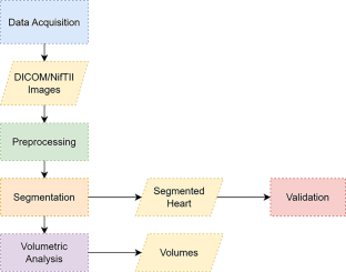
This work addresses the above problems. A new, image-driven, fast, and fully automatic segmentation method was developed to segment the heart from CT images using a processing pipeline of adaptive median filter, multi-level thresholding, active contours, mathematical morphology, and the knowledge of human anatomy to delineate the regions of interest.
Results
The algorithm proposed is simple to implement and validate and requires no human intervention. The method is tested on the 'Image CHD' DICOM images (multi-centre, clinically approved single-phase de-identified images), and the results obtained were validated against the ground truths provided with the dataset. The results show an average Dice score, Jaccard score, and Hausdorff distance of 0.866, 0.776, and 33.29 mm, respectively, for the segmentation of the heart's chambers, aorta, and blood vessels. The results and the ground truths were compared using Bland-Altmon plots.
Conclusion
The heart was correctly segmented from the CT images using the proposed method. Further this segmentation technique can be used to develop AI based solutions for segmentation.
期刊介绍:
Cardiovascular Engineering and Technology is a journal publishing the spectrum of basic to translational research in all aspects of cardiovascular physiology and medical treatment. It is the forum for academic and industrial investigators to disseminate research that utilizes engineering principles and methods to advance fundamental knowledge and technological solutions related to the cardiovascular system. Manuscripts spanning from subcellular to systems level topics are invited, including but not limited to implantable medical devices, hemodynamics and tissue biomechanics, functional imaging, surgical devices, electrophysiology, tissue engineering and regenerative medicine, diagnostic instruments, transport and delivery of biologics, and sensors. In addition to manuscripts describing the original publication of research, manuscripts reviewing developments in these topics or their state-of-art are also invited.
 求助内容:
求助内容: 应助结果提醒方式:


