Susan Wopat, Priyom Adhyapok, Bijoy Daga, Janice M. Crawford, James Norman, Jennifer Bagwell, Brianna Peskin, Indrasen Magre, Stephanie M. Fogerson, Daniel S. Levic, Stefano Di Talia, Daniel P. Kiehart, Patrick Charbonneau, Michel Bagnat
{"title":"Notochord segmentation in zebrafish controlled by iterative mechanical signaling","authors":"Susan Wopat, Priyom Adhyapok, Bijoy Daga, Janice M. Crawford, James Norman, Jennifer Bagwell, Brianna Peskin, Indrasen Magre, Stephanie M. Fogerson, Daniel S. Levic, Stefano Di Talia, Daniel P. Kiehart, Patrick Charbonneau, Michel Bagnat","doi":"10.1016/j.devcel.2024.04.013","DOIUrl":null,"url":null,"abstract":"<p>In bony fishes, patterning of the vertebral column, or spine, is guided by a metameric blueprint established in the notochord sheath. Notochord segmentation begins days after somitogenesis concludes and can occur in its absence. However, somite patterning defects lead to imprecise notochord segmentation, suggesting that these processes are linked. Here, we identify that interactions between the notochord and the axial musculature ensure precise spatiotemporal segmentation of the zebrafish spine. We demonstrate that myoseptum-notochord linkages drive notochord segment initiation by locally deforming the notochord extracellular matrix and recruiting focal adhesion machinery at these contact points. Irregular somite patterning alters this mechanical signaling, causing non-sequential and dysmorphic notochord segmentation, leading to altered spine development. Using a model that captures myoseptum-notochord interactions, we find that a fixed spatial interval is critical for driving sequential segment initiation. Thus, mechanical coupling of axial tissues facilitates spatiotemporal spine patterning.</p>","PeriodicalId":11157,"journal":{"name":"Developmental cell","volume":null,"pages":null},"PeriodicalIF":10.7000,"publicationDate":"2024-05-01","publicationTypes":"Journal Article","fieldsOfStudy":null,"isOpenAccess":false,"openAccessPdf":"","citationCount":"0","resultStr":null,"platform":"Semanticscholar","paperid":null,"PeriodicalName":"Developmental cell","FirstCategoryId":"99","ListUrlMain":"https://doi.org/10.1016/j.devcel.2024.04.013","RegionNum":1,"RegionCategory":"生物学","ArticlePicture":[],"TitleCN":null,"AbstractTextCN":null,"PMCID":null,"EPubDate":"","PubModel":"","JCR":"Q1","JCRName":"CELL BIOLOGY","Score":null,"Total":0}
引用次数: 0
Abstract
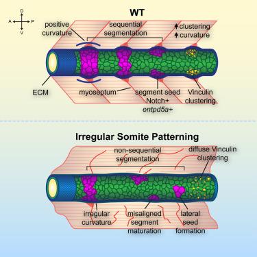
In bony fishes, patterning of the vertebral column, or spine, is guided by a metameric blueprint established in the notochord sheath. Notochord segmentation begins days after somitogenesis concludes and can occur in its absence. However, somite patterning defects lead to imprecise notochord segmentation, suggesting that these processes are linked. Here, we identify that interactions between the notochord and the axial musculature ensure precise spatiotemporal segmentation of the zebrafish spine. We demonstrate that myoseptum-notochord linkages drive notochord segment initiation by locally deforming the notochord extracellular matrix and recruiting focal adhesion machinery at these contact points. Irregular somite patterning alters this mechanical signaling, causing non-sequential and dysmorphic notochord segmentation, leading to altered spine development. Using a model that captures myoseptum-notochord interactions, we find that a fixed spatial interval is critical for driving sequential segment initiation. Thus, mechanical coupling of axial tissues facilitates spatiotemporal spine patterning.
期刊介绍:
Developmental Cell, established in 2001, is a comprehensive journal that explores a wide range of topics in cell and developmental biology. Our publication encompasses work across various disciplines within biology, with a particular emphasis on investigating the intersections between cell biology, developmental biology, and other related fields. Our primary objective is to present research conducted through a cell biological perspective, addressing the essential mechanisms governing cell function, cellular interactions, and responses to the environment. Moreover, we focus on understanding the collective behavior of cells, culminating in the formation of tissues, organs, and whole organisms, while also investigating the consequences of any malfunctions in these intricate processes.
 求助内容:
求助内容: 应助结果提醒方式:


