Viral entry and translation in brain endothelia provoke influenza-associated encephalopathy
Abstract
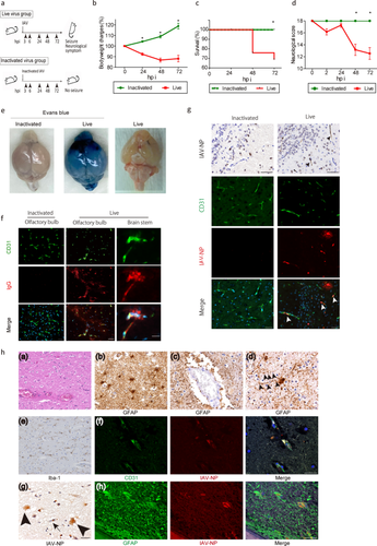
Influenza-associated encephalopathy (IAE) is extremely acute in onset, with high lethality and morbidity within a few days, while the direct pathogenesis by influenza virus in this acute phase in the brain is largely unknown. Here we show that influenza virus enters into the cerebral endothelium and thereby induces IAE. Three-weeks-old young mice were inoculated with influenza A virus (IAV). Physical and neurological scores were recorded and temporal-spatial analyses of histopathology and viral studies were performed up to 72 h post inoculation. Histopathological examinations were also performed using IAE human autopsy brains. Viral infection, proliferation and pathogenesis were analyzed in cell lines of endothelium and astrocyte. The effects of anti-influenza viral drugs were tested in the cell lines and animal models. Upon intravenous inoculation of IAV in mice, the mice developed encephalopathy with brain edema and pathological lesions represented by micro bleeding and injured astrocytic process (clasmatodendrosis) within 72 h. Histologically, massive deposits of viral nucleoprotein were observed as early as 24 h post infection in the brain endothelial cells of mouse models and the IAE patients. IAV inoculated endothelial cell lines showed deposition of viral proteins and provoked cell death, while IAV scarcely amplified. Inhibition of viral transcription and translation suppressed the endothelial cell death and the lethality of mouse models. These data suggest that the onset of encephalopathy should be induced by cerebral endothelial infection with IAV. Thus, IAV entry into the endothelium, and transcription and/or translation of viral RNA, but not viral proliferation, should be the key pathogenesis of IAE.


 求助内容:
求助内容: 应助结果提醒方式:


