A. V. Selezneva, E. V. Korobko, S. L. Kiselev, Yu. G. Suzdaltseva
{"title":"Expression Profile of Isogenic Early Mesodermal Cells Differentiated from Human Induced Pluripotent Stem Cells","authors":"A. V. Selezneva, E. V. Korobko, S. L. Kiselev, Yu. G. Suzdaltseva","doi":"10.1134/s0022093024020042","DOIUrl":null,"url":null,"abstract":"<h3 data-test=\"abstract-sub-heading\">Abstract</h3><p>Scar formation during normal regeneration of damaged tissue\ncan lead to noticeable cosmetic and functional defects of organs\nand thus significantly affect the quality of life. Meanwhile, fetal\ntissues before the third trimester of pregnancy are known to be\ncapable of complete regeneration with the restoration of the original\narchitecture and functional activity. Understanding the cellular\nand molecular mechanisms of fetal wound regeneration will provide\nthe basis for the development of successful treatments aimed to minimize\nscarring. Mesenchymal stromal cells (MSCs) play an important role\nin tissue repair, since cytokines, chemokines, growth factors and\nextracellular vesicles they secrete are involved in the regulation\nof migration, angiogenesis, synthesis, and remodeling of the extracellular\nmatrix. Mesodermal differentiation of human induced pluripotent\nstem cells (iPSCs) enables to reproduce consecutive stages of embryogenesis\nin vitro and to create isogenic cell models of MSCs, corresponding\nto different stages of human development. Here, we performed a specifically\ndirected, multistage, mesodermal differentiation of iPSCs into isogenic\ncell lines of the primitive streak, lateral and paraxial mesoderm,\nas well as carried out a comparative analysis of their expression\nprofiles. It was shown that the derived cells of the lateral mesoderm\n(LM) and paraxial mesoderm (PM) are precursors for MSCs. The MSCs,\nderived due to differentiation of both LM and PM cells, shared a\nsimilar expression profile of pan-mesodermal markers. A comparative\nanalysis of the functional activity of MSCs and their precursors\nin a pro-inflammatory microenvironment will hopefully provide molecular\ntools for a better insight into the basic mechanisms of fetal tissue\nregeneration and help identify therapeutic targets to minimize scarring\nand pathological processes characterized by excessive fibroplasia.</p>","PeriodicalId":15805,"journal":{"name":"Journal of Evolutionary Biochemistry and Physiology","volume":"23 1","pages":""},"PeriodicalIF":0.5000,"publicationDate":"2024-04-26","publicationTypes":"Journal Article","fieldsOfStudy":null,"isOpenAccess":false,"openAccessPdf":"","citationCount":"0","resultStr":null,"platform":"Semanticscholar","paperid":null,"PeriodicalName":"Journal of Evolutionary Biochemistry and Physiology","FirstCategoryId":"99","ListUrlMain":"https://doi.org/10.1134/s0022093024020042","RegionNum":4,"RegionCategory":"生物学","ArticlePicture":[],"TitleCN":null,"AbstractTextCN":null,"PMCID":null,"EPubDate":"","PubModel":"","JCR":"Q4","JCRName":"BIOCHEMISTRY & MOLECULAR BIOLOGY","Score":null,"Total":0}
引用次数: 0
Abstract
Scar formation during normal regeneration of damaged tissue
can lead to noticeable cosmetic and functional defects of organs
and thus significantly affect the quality of life. Meanwhile, fetal
tissues before the third trimester of pregnancy are known to be
capable of complete regeneration with the restoration of the original
architecture and functional activity. Understanding the cellular
and molecular mechanisms of fetal wound regeneration will provide
the basis for the development of successful treatments aimed to minimize
scarring. Mesenchymal stromal cells (MSCs) play an important role
in tissue repair, since cytokines, chemokines, growth factors and
extracellular vesicles they secrete are involved in the regulation
of migration, angiogenesis, synthesis, and remodeling of the extracellular
matrix. Mesodermal differentiation of human induced pluripotent
stem cells (iPSCs) enables to reproduce consecutive stages of embryogenesis
in vitro and to create isogenic cell models of MSCs, corresponding
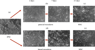
to different stages of human development. Here, we performed a specifically
directed, multistage, mesodermal differentiation of iPSCs into isogenic
cell lines of the primitive streak, lateral and paraxial mesoderm,
as well as carried out a comparative analysis of their expression
profiles. It was shown that the derived cells of the lateral mesoderm
(LM) and paraxial mesoderm (PM) are precursors for MSCs. The MSCs,
derived due to differentiation of both LM and PM cells, shared a
similar expression profile of pan-mesodermal markers. A comparative
analysis of the functional activity of MSCs and their precursors
in a pro-inflammatory microenvironment will hopefully provide molecular
tools for a better insight into the basic mechanisms of fetal tissue
regeneration and help identify therapeutic targets to minimize scarring
and pathological processes characterized by excessive fibroplasia.
期刊介绍:
Journal of Evolutionary Biochemistry and Physiology publishes original experimental and theoretical and review articles related to evolution of the main forms of metabolism in connection with life origin; comparative and ontogenetic physiology and biochemistry, biochemical evolution of animal world; as well as evolution of functions; morphology, pharmacology, pathophysiology and ecological physiology. The journal welcomes manuscripts from all countries in the English or Russian language.
 求助内容:
求助内容: 应助结果提醒方式:


