Facile engineering of aptamer-coupled silk fibroin encapsulated myogenic gold nanocomposites: investigation of antiproliferative activity and apoptosis induction
IF 2 4区 生物学Q3 BIOTECHNOLOGY & APPLIED MICROBIOLOGY
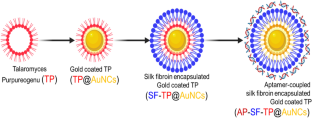
{"title":"Facile engineering of aptamer-coupled silk fibroin encapsulated myogenic gold nanocomposites: investigation of antiproliferative activity and apoptosis induction","authors":"Poorni Kaliyappan Elayappan, Kavitha Kandasamy, Vadivukkarasi Sasikumar, Muruganantham Bharathi, Abdurahman Hajinur Hirad, Abdullah A. Alarfaj, Palanisamy Arulselvan, Ravindran Jaganathan, Rajeswari Ravindran, Jagadeesh Suriyaprakash, Indumathi Thangavelu","doi":"10.1007/s10529-024-03491-2","DOIUrl":null,"url":null,"abstract":"<p>Nanocomposites selectively induce cancer cell death, holding potential for precise liver cancer treatment breakthroughs. This study assessed the cytotoxicity of gold nanocomposites (Au NCs) enclosed within silk fibroin (SF), aptamer (Ap), and the myogenic <i>Talaromyces purpureogenus</i> (TP) against a human liver cancer cell (HepG2). The ultimate product, Ap-SF-TP@Au NCs, results from a three-step process. This process involves the myogenic synthesis of TP@Au NCs derived from TP mycelial extract, encapsulation of SF on TP@Au NCs (SF-TP@Au NCs), and the conjugation of Ap within SF-TP@Au NCs. The synthesized NCs are analyzed by various characteristic techniques. Ap-SF-TP@Au NCs induced potential cell death in HepG2 cells but exhibited no cytotoxicity in non-cancerous cells (NIH3T3). The morphological changes in cells were examined through various biochemical staining methods. Thus, Ap-SF-TP@Au NCs emerge as a promising nanocomposite for treating diverse cancer cells.</p>","PeriodicalId":8929,"journal":{"name":"Biotechnology Letters","volume":null,"pages":null},"PeriodicalIF":2.0000,"publicationDate":"2024-04-27","publicationTypes":"Journal Article","fieldsOfStudy":null,"isOpenAccess":false,"openAccessPdf":"","citationCount":"0","resultStr":null,"platform":"Semanticscholar","paperid":null,"PeriodicalName":"Biotechnology Letters","FirstCategoryId":"5","ListUrlMain":"https://doi.org/10.1007/s10529-024-03491-2","RegionNum":4,"RegionCategory":"生物学","ArticlePicture":[],"TitleCN":null,"AbstractTextCN":null,"PMCID":null,"EPubDate":"","PubModel":"","JCR":"Q3","JCRName":"BIOTECHNOLOGY & APPLIED MICROBIOLOGY","Score":null,"Total":0}
引用次数: 0
Abstract
Nanocomposites selectively induce cancer cell death, holding potential for precise liver cancer treatment breakthroughs. This study assessed the cytotoxicity of gold nanocomposites (Au NCs) enclosed within silk fibroin (SF), aptamer (Ap), and the myogenic Talaromyces purpureogenus (TP) against a human liver cancer cell (HepG2). The ultimate product, Ap-SF-TP@Au NCs, results from a three-step process. This process involves the myogenic synthesis of TP@Au NCs derived from TP mycelial extract, encapsulation of SF on TP@Au NCs (SF-TP@Au NCs), and the conjugation of Ap within SF-TP@Au NCs. The synthesized NCs are analyzed by various characteristic techniques. Ap-SF-TP@Au NCs induced potential cell death in HepG2 cells but exhibited no cytotoxicity in non-cancerous cells (NIH3T3). The morphological changes in cells were examined through various biochemical staining methods. Thus, Ap-SF-TP@Au NCs emerge as a promising nanocomposite for treating diverse cancer cells.
期刊介绍:
Biotechnology Letters is the world’s leading rapid-publication primary journal dedicated to biotechnology as a whole – that is to topics relating to actual or potential applications of biological reactions affected by microbial, plant or animal cells and biocatalysts derived from them.
All relevant aspects of molecular biology, genetics and cell biochemistry, of process and reactor design, of pre- and post-treatment steps, and of manufacturing or service operations are therefore included.
Contributions from industrial and academic laboratories are equally welcome. We also welcome contributions covering biotechnological aspects of regenerative medicine and biomaterials and also cancer biotechnology. Criteria for the acceptance of papers relate to our aim of publishing useful and informative results that will be of value to other workers in related fields.
The emphasis is very much on novelty and immediacy in order to justify rapid publication of authors’ results. It should be noted, however, that we do not normally publish papers (but this is not absolute) that deal with unidentified consortia of microorganisms (e.g. as in activated sludge) as these results may not be easily reproducible in other laboratories.
Papers describing the isolation and identification of microorganisms are not regarded as appropriate but such information can be appended as supporting information to a paper. Papers dealing with simple process development are usually considered to lack sufficient novelty or interest to warrant publication.
 求助内容:
求助内容: 应助结果提醒方式:


