Aarshi N. Singh, Justin B Nice, Meishan Wu, Angela C. Brown and Nathan J. Wittenberg*,
{"title":"Multivariate Analysis of Individual Bacterial Outer Membrane Vesicles Using Fluorescence Microscopy","authors":"Aarshi N. Singh, Justin B Nice, Meishan Wu, Angela C. Brown and Nathan J. Wittenberg*, ","doi":"10.1021/cbmi.4c00014","DOIUrl":null,"url":null,"abstract":"<p >Gram-negative bacteria produce outer membrane vesicles (OMVs) that play a critical role in cell–cell communication and virulence. OMVs have emerged as promising therapeutic agents for various biological applications such as vaccines and targeted drug delivery. However, the full potential of OMVs is currently constrained by inherent heterogeneities, such as size and cargo differences, and traditional ensemble assays are limited in their ability to reveal OMV heterogeneity. To overcome this issue, we devised an innovative approach enabling the identification of various characteristics of individual OMVs. This method, employing fluorescence microscopy, facilitates the detection of variations in size and surface markers. To demonstrate our method, we utilize the oral bacterium <i>Aggregatibacter actinomycetemcomitans (A. actinomycetemcomitans)</i> which produces OMVs with a bimodal size distribution. As part of its virulence, <i>A. actinomycetemcomitans</i> secretes leukotoxin (LtxA) in two forms: soluble and surface associated with the OMVs. We observed a correlation between the size and toxin presence where larger OMVs were much more likely to possess LtxA compared to the smaller OMVs. In addition, we noted that, among the smallest OMVs (<100 nm diameter), the fractions that are toxin positive range from 0 to 30%, while the largest OMVs (>200 nm diameter) are between 70 and 100% toxin positive.</p>","PeriodicalId":53181,"journal":{"name":"Chemical & Biomedical Imaging","volume":"2 5","pages":"352–361"},"PeriodicalIF":0.0000,"publicationDate":"2024-04-19","publicationTypes":"Journal Article","fieldsOfStudy":null,"isOpenAccess":false,"openAccessPdf":"https://pubs.acs.org/doi/epdf/10.1021/cbmi.4c00014","citationCount":"0","resultStr":null,"platform":"Semanticscholar","paperid":null,"PeriodicalName":"Chemical & Biomedical Imaging","FirstCategoryId":"1085","ListUrlMain":"https://pubs.acs.org/doi/10.1021/cbmi.4c00014","RegionNum":0,"RegionCategory":null,"ArticlePicture":[],"TitleCN":null,"AbstractTextCN":null,"PMCID":null,"EPubDate":"","PubModel":"","JCR":"","JCRName":"","Score":null,"Total":0}
引用次数: 0
Abstract
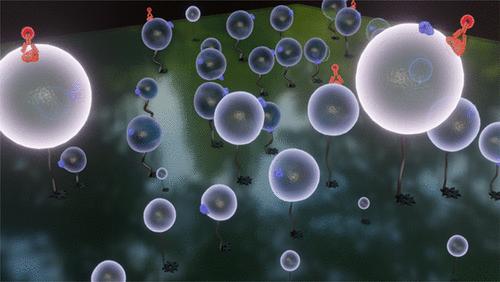
Gram-negative bacteria produce outer membrane vesicles (OMVs) that play a critical role in cell–cell communication and virulence. OMVs have emerged as promising therapeutic agents for various biological applications such as vaccines and targeted drug delivery. However, the full potential of OMVs is currently constrained by inherent heterogeneities, such as size and cargo differences, and traditional ensemble assays are limited in their ability to reveal OMV heterogeneity. To overcome this issue, we devised an innovative approach enabling the identification of various characteristics of individual OMVs. This method, employing fluorescence microscopy, facilitates the detection of variations in size and surface markers. To demonstrate our method, we utilize the oral bacterium Aggregatibacter actinomycetemcomitans (A. actinomycetemcomitans) which produces OMVs with a bimodal size distribution. As part of its virulence, A. actinomycetemcomitans secretes leukotoxin (LtxA) in two forms: soluble and surface associated with the OMVs. We observed a correlation between the size and toxin presence where larger OMVs were much more likely to possess LtxA compared to the smaller OMVs. In addition, we noted that, among the smallest OMVs (<100 nm diameter), the fractions that are toxin positive range from 0 to 30%, while the largest OMVs (>200 nm diameter) are between 70 and 100% toxin positive.
期刊介绍:
Chemical & Biomedical Imaging is a peer-reviewed open access journal devoted to the publication of cutting-edge research papers on all aspects of chemical and biomedical imaging. This interdisciplinary field sits at the intersection of chemistry physics biology materials engineering and medicine. The journal aims to bring together researchers from across these disciplines to address cutting-edge challenges of fundamental research and applications.Topics of particular interest include but are not limited to:Imaging of processes and reactionsImaging of nanoscale microscale and mesoscale materialsImaging of biological interactions and interfacesSingle-molecule and cellular imagingWhole-organ and whole-body imagingMolecular imaging probes and contrast agentsBioluminescence chemiluminescence and electrochemiluminescence imagingNanophotonics and imagingChemical tools for new imaging modalitiesChemical and imaging techniques in diagnosis and therapyImaging-guided drug deliveryAI and machine learning assisted imaging
 求助内容:
求助内容: 应助结果提醒方式:


