Fatma Ben Trad, Jérôme Delacotte, Frédéric Lemaître, Manon Guille-Collignon, Stéphane Arbault, Neso Sojic, Eric Labbé and Olivier Buriez
{"title":"Shadow electrochemiluminescence imaging of giant liposomes opening at polarized electrodes†","authors":"Fatma Ben Trad, Jérôme Delacotte, Frédéric Lemaître, Manon Guille-Collignon, Stéphane Arbault, Neso Sojic, Eric Labbé and Olivier Buriez","doi":"10.1039/D4AN00470A","DOIUrl":null,"url":null,"abstract":"<p >In this work, the release of giant liposome (∼100 μm in diameter) content was imaged by shadow electrochemiluminescence (ECL) microscopy. Giant unilamellar liposomes were pre-loaded with a sucrose solution and allowed to sediment at an ITO electrode surface immersed in a solution containing a luminophore ([Ru(bpy)<small><sub>3</sub></small>]<small><sup>2+</sup></small>) and a sacrificial co-reactant (tri-<em>n</em>-propylamine). Upon polarization, the electrode exhibited illumination over its entire surface thanks to the oxidation of ECL reagents. However, as soon as liposomes reached the electrode surface, dark spots appeared and then spread over time on the surface. This observation reflected a blockage of the electrode surface at the contact point between the liposome and the electrode surface, followed by the dilution of ECL reagents after the rupture of the liposome membrane and release of its internal ECL-inactive solution. Interestingly, ECL reappeared in areas where it initially faded, indicating back-diffusion of ECL reagents towards the previously diluted area and thus confirming liposome permeabilization. The whole process was analyzed qualitatively and quantitatively within the defined region of interest. Two mass transport regimes were identified: a gravity-driven spreading process when the liposome releases its content leading to ECL vanishing and a diffusive regime when ECL recovers. The reported shadow ECL microscopy should find promising applications for the imaging of transient events such as molecular species released by artificial or biological vesicles.</p>","PeriodicalId":63,"journal":{"name":"Analyst","volume":" 12","pages":" 3317-3324"},"PeriodicalIF":3.3000,"publicationDate":"2024-04-23","publicationTypes":"Journal Article","fieldsOfStudy":null,"isOpenAccess":false,"openAccessPdf":"","citationCount":"0","resultStr":null,"platform":"Semanticscholar","paperid":null,"PeriodicalName":"Analyst","FirstCategoryId":"92","ListUrlMain":"https://pubs.rsc.org/en/content/articlelanding/2024/an/d4an00470a","RegionNum":3,"RegionCategory":"化学","ArticlePicture":[],"TitleCN":null,"AbstractTextCN":null,"PMCID":null,"EPubDate":"","PubModel":"","JCR":"Q2","JCRName":"CHEMISTRY, ANALYTICAL","Score":null,"Total":0}
引用次数: 0
Abstract
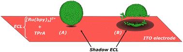
In this work, the release of giant liposome (∼100 μm in diameter) content was imaged by shadow electrochemiluminescence (ECL) microscopy. Giant unilamellar liposomes were pre-loaded with a sucrose solution and allowed to sediment at an ITO electrode surface immersed in a solution containing a luminophore ([Ru(bpy)3]2+) and a sacrificial co-reactant (tri-n-propylamine). Upon polarization, the electrode exhibited illumination over its entire surface thanks to the oxidation of ECL reagents. However, as soon as liposomes reached the electrode surface, dark spots appeared and then spread over time on the surface. This observation reflected a blockage of the electrode surface at the contact point between the liposome and the electrode surface, followed by the dilution of ECL reagents after the rupture of the liposome membrane and release of its internal ECL-inactive solution. Interestingly, ECL reappeared in areas where it initially faded, indicating back-diffusion of ECL reagents towards the previously diluted area and thus confirming liposome permeabilization. The whole process was analyzed qualitatively and quantitatively within the defined region of interest. Two mass transport regimes were identified: a gravity-driven spreading process when the liposome releases its content leading to ECL vanishing and a diffusive regime when ECL recovers. The reported shadow ECL microscopy should find promising applications for the imaging of transient events such as molecular species released by artificial or biological vesicles.
 求助内容:
求助内容: 应助结果提醒方式:


