Damian Dudkiewicz, Maciej Lis, Artem Yakovliev, Jakub Hołda, Filip Bolechała, Marcin Strona, Paweł Kopacz, Mateusz K. Hołda
{"title":"Aortic root morphometry revisited—Clinical implications for aortic valve interventions","authors":"Damian Dudkiewicz, Maciej Lis, Artem Yakovliev, Jakub Hołda, Filip Bolechała, Marcin Strona, Paweł Kopacz, Mateusz K. Hołda","doi":"10.1002/ca.24165","DOIUrl":null,"url":null,"abstract":"<p>The complex anatomy of the aortic root is of great importance for many surgical and transcatheter cardiac procedures. Therefore, the aim of this study was to provide a comprehensive morphological description of the nondiseased aortic root. We morphometrically examined 200 autopsied human adult hearts (22.0% females, 47.9 ± 17.7 years). A meticulous macroscopic analysis of aortic root anatomy was performed. The largest cross-section area of the aortic root was observed in coaptation center plane (653.9 ± 196.5 mm<sup>2</sup>), followed by tubular plane (427.7 ± 168.0 mm<sup>2</sup>) and basal ring (362.7 ± 159.1 mm<sup>2</sup>) (<i>p</i> < 0.001). The right coronary sinus was the largest (area: 234.3 ± 85.0 mm<sup>2</sup>), followed by noncoronary sinus (218.7 ± 74.8 mm<sup>2</sup>) and left coronary sinus (201.2 ± 78.08 mm<sup>2</sup>). The noncoronary sinus was the deepest, followed by right and left coronary sinus (16.4 ± 3.2 vs. 15.9 ± 3.1 vs. 14.9 ± 2.9 mm, <i>p</i> < 0.001). In 68.5% of hearts, the coaptation center was located near the aortic geometric center. The left coronary ostium was located 15.6 ± 3.8 mm above sinus bottom (within the sinus in 91.5% and above sinutubular junction in 8.5%), while for right coronary ostium, it was 16.2 ± 3.5 mm above (83.5% within sinus and 16.5% above). In general, males exhibited larger aortic valve dimensions than females. A multiple forward stepwise regression model showed that anthropometric variables might predict the size of coaptation center plane (age, sex, and heart weight; <i>R</i><sup>2</sup> = 31.8%), tubular plane (age and sex; <i>R</i><sup>2</sup> = 25.6%), and basal ring (age and sex; <i>R</i><sup>2</sup> = 16.9%). In conclusion, this study presents a comprehensive analysis of aortic-root morphometry and provides a platform for further research into the intricate interplay between structure and function of the aortic root.</p>","PeriodicalId":50687,"journal":{"name":"Clinical Anatomy","volume":"37 7","pages":"719-729"},"PeriodicalIF":2.3000,"publicationDate":"2024-04-17","publicationTypes":"Journal Article","fieldsOfStudy":null,"isOpenAccess":false,"openAccessPdf":"https://onlinelibrary.wiley.com/doi/epdf/10.1002/ca.24165","citationCount":"0","resultStr":null,"platform":"Semanticscholar","paperid":null,"PeriodicalName":"Clinical Anatomy","FirstCategoryId":"3","ListUrlMain":"https://onlinelibrary.wiley.com/doi/10.1002/ca.24165","RegionNum":4,"RegionCategory":"医学","ArticlePicture":[],"TitleCN":null,"AbstractTextCN":null,"PMCID":null,"EPubDate":"","PubModel":"","JCR":"Q1","JCRName":"ANATOMY & MORPHOLOGY","Score":null,"Total":0}
引用次数: 0
Abstract
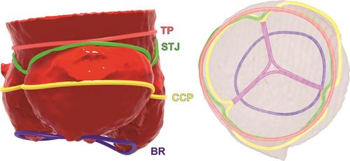
The complex anatomy of the aortic root is of great importance for many surgical and transcatheter cardiac procedures. Therefore, the aim of this study was to provide a comprehensive morphological description of the nondiseased aortic root. We morphometrically examined 200 autopsied human adult hearts (22.0% females, 47.9 ± 17.7 years). A meticulous macroscopic analysis of aortic root anatomy was performed. The largest cross-section area of the aortic root was observed in coaptation center plane (653.9 ± 196.5 mm2), followed by tubular plane (427.7 ± 168.0 mm2) and basal ring (362.7 ± 159.1 mm2) (p < 0.001). The right coronary sinus was the largest (area: 234.3 ± 85.0 mm2), followed by noncoronary sinus (218.7 ± 74.8 mm2) and left coronary sinus (201.2 ± 78.08 mm2). The noncoronary sinus was the deepest, followed by right and left coronary sinus (16.4 ± 3.2 vs. 15.9 ± 3.1 vs. 14.9 ± 2.9 mm, p < 0.001). In 68.5% of hearts, the coaptation center was located near the aortic geometric center. The left coronary ostium was located 15.6 ± 3.8 mm above sinus bottom (within the sinus in 91.5% and above sinutubular junction in 8.5%), while for right coronary ostium, it was 16.2 ± 3.5 mm above (83.5% within sinus and 16.5% above). In general, males exhibited larger aortic valve dimensions than females. A multiple forward stepwise regression model showed that anthropometric variables might predict the size of coaptation center plane (age, sex, and heart weight; R2 = 31.8%), tubular plane (age and sex; R2 = 25.6%), and basal ring (age and sex; R2 = 16.9%). In conclusion, this study presents a comprehensive analysis of aortic-root morphometry and provides a platform for further research into the intricate interplay between structure and function of the aortic root.
期刊介绍:
Clinical Anatomy is the Official Journal of the American Association of Clinical Anatomists and the British Association of Clinical Anatomists. The goal of Clinical Anatomy is to provide a medium for the exchange of current information between anatomists and clinicians. This journal embraces anatomy in all its aspects as applied to medical practice. Furthermore, the journal assists physicians and other health care providers in keeping abreast of new methodologies for patient management and informs educators of new developments in clinical anatomy and teaching techniques. Clinical Anatomy publishes original and review articles of scientific, clinical, and educational interest. Papers covering the application of anatomic principles to the solution of clinical problems and/or the application of clinical observations to expand anatomic knowledge are welcomed.

 求助内容:
求助内容: 应助结果提醒方式:


