Gut—brain barrier dysfunction bridge autistic-like behavior in mouse model of maternal separation stress: A behavioral, histopathological, and molecular study
{"title":"Gut—brain barrier dysfunction bridge autistic-like behavior in mouse model of maternal separation stress: A behavioral, histopathological, and molecular study","authors":"Negin Rowshan, Maryam Anjomshoa, Anahita Farahzad, Elham Bijad, Hossein Amini-Khoei","doi":"10.1002/jdn.10329","DOIUrl":null,"url":null,"abstract":"<p>Autism spectrum disorder (ASD) is a fast-growing neurodevelopmental disorder throughout the world. Experiencing early life stresses (ELS) like maternal separation (MS) is associated with autistic-like behaviors. It has been proposed that disturbance in the gut–brain axis-mediated psychiatric disorders following MS. The role of disruption in the integrity of gut–brain barrier in ASD remains unclear. Addressing this knowledge gap, in this study we aimed to investigate role of the gut–brain barrier integrity in mediating autistic-like behaviors in mouse models of MS stress. To do this, mice neonates are separated daily from their mothers from postnatal day (PND) 2 to PND 14 for 3 hours. During PND58–60, behavioral tests related to autistic-like behaviors including three-chamber sociability, shuttle box, and resident-intruder tests were performed. Then, prefrontal cortex (PFC), hippocampus, and colon samples were dissected out for histopathological and molecular evaluations. Results showed that MS is associated with impaired sociability and social preference indexes, aggressive behaviors, and impaired passive avoidance memory. The gene expression of CLDN1 decreased in the colon, and the gene expression of CLDN5, CLDN12, and MMP9 increased in the PFC of the MS mice. MS is associated with decrease in the diameter of CA1 and CA3 areas of the hippocampus. In addition, MS led to histopathological changes in the colon. We concluded that, probably, disturbance in the gut–brain barrier integrities mediated the autistic-like behavior in MS stress in mice.</p>","PeriodicalId":13914,"journal":{"name":"International Journal of Developmental Neuroscience","volume":"84 4","pages":"314-327"},"PeriodicalIF":1.7000,"publicationDate":"2024-04-07","publicationTypes":"Journal Article","fieldsOfStudy":null,"isOpenAccess":false,"openAccessPdf":"","citationCount":"0","resultStr":null,"platform":"Semanticscholar","paperid":null,"PeriodicalName":"International Journal of Developmental Neuroscience","FirstCategoryId":"3","ListUrlMain":"https://onlinelibrary.wiley.com/doi/10.1002/jdn.10329","RegionNum":4,"RegionCategory":"医学","ArticlePicture":[],"TitleCN":null,"AbstractTextCN":null,"PMCID":null,"EPubDate":"","PubModel":"","JCR":"Q3","JCRName":"DEVELOPMENTAL BIOLOGY","Score":null,"Total":0}
引用次数: 0
Abstract
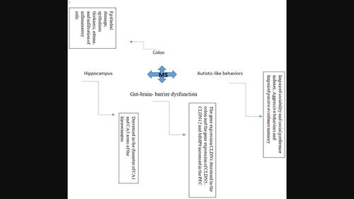
Autism spectrum disorder (ASD) is a fast-growing neurodevelopmental disorder throughout the world. Experiencing early life stresses (ELS) like maternal separation (MS) is associated with autistic-like behaviors. It has been proposed that disturbance in the gut–brain axis-mediated psychiatric disorders following MS. The role of disruption in the integrity of gut–brain barrier in ASD remains unclear. Addressing this knowledge gap, in this study we aimed to investigate role of the gut–brain barrier integrity in mediating autistic-like behaviors in mouse models of MS stress. To do this, mice neonates are separated daily from their mothers from postnatal day (PND) 2 to PND 14 for 3 hours. During PND58–60, behavioral tests related to autistic-like behaviors including three-chamber sociability, shuttle box, and resident-intruder tests were performed. Then, prefrontal cortex (PFC), hippocampus, and colon samples were dissected out for histopathological and molecular evaluations. Results showed that MS is associated with impaired sociability and social preference indexes, aggressive behaviors, and impaired passive avoidance memory. The gene expression of CLDN1 decreased in the colon, and the gene expression of CLDN5, CLDN12, and MMP9 increased in the PFC of the MS mice. MS is associated with decrease in the diameter of CA1 and CA3 areas of the hippocampus. In addition, MS led to histopathological changes in the colon. We concluded that, probably, disturbance in the gut–brain barrier integrities mediated the autistic-like behavior in MS stress in mice.
期刊介绍:
International Journal of Developmental Neuroscience publishes original research articles and critical review papers on all fundamental and clinical aspects of nervous system development, renewal and regeneration, as well as on the effects of genetic and environmental perturbations of brain development and homeostasis leading to neurodevelopmental disorders and neurological conditions. Studies describing the involvement of stem cells in nervous system maintenance and disease (including brain tumours), stem cell-based approaches for the investigation of neurodegenerative diseases, roles of neuroinflammation in development and disease, and neuroevolution are also encouraged. Investigations using molecular, cellular, physiological, genetic and epigenetic approaches in model systems ranging from simple invertebrates to human iPSC-based 2D and 3D models are encouraged, as are studies using experimental models that provide behavioural or evolutionary insights. The journal also publishes Special Issues dealing with topics at the cutting edge of research edited by Guest Editors appointed by the Editor in Chief. A major aim of the journal is to facilitate the transfer of fundamental studies of nervous system development, maintenance, and disease to clinical applications. The journal thus intends to disseminate valuable information for both biologists and physicians. International Journal of Developmental Neuroscience is owned and supported by The International Society for Developmental Neuroscience (ISDN), an organization of scientists interested in advancing developmental neuroscience research in the broadest sense.
 求助内容:
求助内容: 应助结果提醒方式:


