Md Nasful Huda Prince, Benjamin Garcia, Cory Henn, Yating Yi, Etsuo A. Susaki, Yuki Watakabe, Tomomi Nemoto, Keith A. Lidke, Hu Zhao, Irene Salinas Remiro, Sheng Liu, Tonmoy Chakraborty
{"title":"Signal improved ultra-fast light-sheet microscope for large tissue imaging","authors":"Md Nasful Huda Prince, Benjamin Garcia, Cory Henn, Yating Yi, Etsuo A. Susaki, Yuki Watakabe, Tomomi Nemoto, Keith A. Lidke, Hu Zhao, Irene Salinas Remiro, Sheng Liu, Tonmoy Chakraborty","doi":"10.1038/s44172-024-00205-4","DOIUrl":null,"url":null,"abstract":"Axially swept light-sheet microscope in conjunction with tissue clearing enables three-dimensional morphological investigation of millimeter-scaled tissues at isotropic sub-micron resolution. However, these microscopes suffer from low detection signal and slow imaging speed. Here we report a simple and efficient imaging platform that employs precise control of two fixed distant light-sheet foci for axial sweeping. This enables full field of view imaging at 40 frames per second, a four-fold improvement over the current state-of-the-art. In addition, in a particular frame rate, our method doubles the signal compared to the existing techniques. To augment the overall imaging performance, we also developed a deep learning based tissue information classifier that enables faster determination of tissue boundary. We demonstrated the performance of our imaging platform on various cleared tissue samples and delineated its robustness over a wide range of clearing protocols. Md Nasful Huda Prince and colleagues propose a tissue imaging system with isotropic sub-micron resolution. The method intelligently delineates tissue borders and captures images faster and with enhanced signal quality.","PeriodicalId":72644,"journal":{"name":"Communications engineering","volume":null,"pages":null},"PeriodicalIF":0.0000,"publicationDate":"2024-04-02","publicationTypes":"Journal Article","fieldsOfStudy":null,"isOpenAccess":false,"openAccessPdf":"https://www.nature.com/articles/s44172-024-00205-4.pdf","citationCount":"0","resultStr":null,"platform":"Semanticscholar","paperid":null,"PeriodicalName":"Communications engineering","FirstCategoryId":"1085","ListUrlMain":"https://www.nature.com/articles/s44172-024-00205-4","RegionNum":0,"RegionCategory":null,"ArticlePicture":[],"TitleCN":null,"AbstractTextCN":null,"PMCID":null,"EPubDate":"","PubModel":"","JCR":"","JCRName":"","Score":null,"Total":0}
引用次数: 0
Abstract
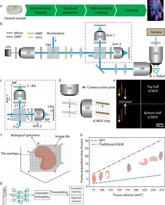
Axially swept light-sheet microscope in conjunction with tissue clearing enables three-dimensional morphological investigation of millimeter-scaled tissues at isotropic sub-micron resolution. However, these microscopes suffer from low detection signal and slow imaging speed. Here we report a simple and efficient imaging platform that employs precise control of two fixed distant light-sheet foci for axial sweeping. This enables full field of view imaging at 40 frames per second, a four-fold improvement over the current state-of-the-art. In addition, in a particular frame rate, our method doubles the signal compared to the existing techniques. To augment the overall imaging performance, we also developed a deep learning based tissue information classifier that enables faster determination of tissue boundary. We demonstrated the performance of our imaging platform on various cleared tissue samples and delineated its robustness over a wide range of clearing protocols. Md Nasful Huda Prince and colleagues propose a tissue imaging system with isotropic sub-micron resolution. The method intelligently delineates tissue borders and captures images faster and with enhanced signal quality.

 求助内容:
求助内容: 应助结果提醒方式:


