Mads Eg Andersen , Ulf Andersen , Lars Wiking , Jan Trige Rasmussen , Milena Corredig , Sandra Beyer Gregersen
{"title":"The exploration of milk fat crystallization in milk fat globules by confocal Raman microscopy","authors":"Mads Eg Andersen , Ulf Andersen , Lars Wiking , Jan Trige Rasmussen , Milena Corredig , Sandra Beyer Gregersen","doi":"10.1016/j.foostr.2024.100372","DOIUrl":null,"url":null,"abstract":"<div><p>Crystallization behavior within oil-in-water emulsion is a key factor for the properties and stability of many food products. Confocal Raman microscopy is a promising method to study such complex structures in situ. This study aimed at evaluating the feasibility of confocal Raman microscopy for visualizing milk fat crystallization and establishing a reliable data acquisition and processing methodology. Milk fat globules from raw milk were fixated in an agarose gel and crystallized at different temperatures. Confocal Raman microscopy was applied to collect two-dimensional area scans and supporting images were obtained by polarized light microscopy. The results revealed differences in lipid characteristics, crystal formation, and spatial distribution as a result of crystallization. Specific C-C stretching vibrations at 1063, 1083, and 1125 cm-1 were found to indicate lipid chain mobility and provide quantitative information on crystallinity. Additionally, the study successfully identified the triple-layered milk fat globule membrane. Different approaches for processing spectroscopic data were compared, emphasizing the importance of proper data handling. This novel spectroscopy imaging approach has significant potential in enhancing our understanding of structural heterogeneities of crystallized structures within complex food matrices.</p></div>","PeriodicalId":48640,"journal":{"name":"Food Structure-Netherlands","volume":"40 ","pages":"Article 100372"},"PeriodicalIF":5.9000,"publicationDate":"2024-04-01","publicationTypes":"Journal Article","fieldsOfStudy":null,"isOpenAccess":false,"openAccessPdf":"https://www.sciencedirect.com/science/article/pii/S221332912400008X/pdfft?md5=2cbd530d41e69e38aa3d86079fceb5c5&pid=1-s2.0-S221332912400008X-main.pdf","citationCount":"0","resultStr":null,"platform":"Semanticscholar","paperid":null,"PeriodicalName":"Food Structure-Netherlands","FirstCategoryId":"97","ListUrlMain":"https://www.sciencedirect.com/science/article/pii/S221332912400008X","RegionNum":3,"RegionCategory":"农林科学","ArticlePicture":[],"TitleCN":null,"AbstractTextCN":null,"PMCID":null,"EPubDate":"","PubModel":"","JCR":"Q1","JCRName":"FOOD SCIENCE & TECHNOLOGY","Score":null,"Total":0}
引用次数: 0
Abstract
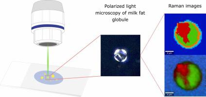
Crystallization behavior within oil-in-water emulsion is a key factor for the properties and stability of many food products. Confocal Raman microscopy is a promising method to study such complex structures in situ. This study aimed at evaluating the feasibility of confocal Raman microscopy for visualizing milk fat crystallization and establishing a reliable data acquisition and processing methodology. Milk fat globules from raw milk were fixated in an agarose gel and crystallized at different temperatures. Confocal Raman microscopy was applied to collect two-dimensional area scans and supporting images were obtained by polarized light microscopy. The results revealed differences in lipid characteristics, crystal formation, and spatial distribution as a result of crystallization. Specific C-C stretching vibrations at 1063, 1083, and 1125 cm-1 were found to indicate lipid chain mobility and provide quantitative information on crystallinity. Additionally, the study successfully identified the triple-layered milk fat globule membrane. Different approaches for processing spectroscopic data were compared, emphasizing the importance of proper data handling. This novel spectroscopy imaging approach has significant potential in enhancing our understanding of structural heterogeneities of crystallized structures within complex food matrices.
期刊介绍:
Food Structure is the premier international forum devoted to the publication of high-quality original research on food structure. The focus of this journal is on food structure in the context of its relationship with molecular composition, processing and macroscopic properties (e.g., shelf stability, sensory properties, etc.). Manuscripts that only report qualitative findings and micrographs and that lack sound hypothesis-driven, quantitative structure-function research are not accepted. Significance of the research findings for the food science community and/or industry must also be highlighted.
 求助内容:
求助内容: 应助结果提醒方式:


