Nondestructive, longitudinal, 3D oxygen imaging of cells in a multi-well plate using pulse electron paramagnetic resonance imaging

Safa Hameed, Navin Viswakarma, Greta Babakhanova, Carl G. Simon Jr., Boris Epel, Mrignayani Kotecha
{"title":"Nondestructive, longitudinal, 3D oxygen imaging of cells in a multi-well plate using pulse electron paramagnetic resonance imaging","authors":"Safa Hameed, Navin Viswakarma, Greta Babakhanova, Carl G. Simon Jr., Boris Epel, Mrignayani Kotecha","doi":"10.1038/s44303-024-00013-7","DOIUrl":null,"url":null,"abstract":"The use of oxygen by cells is an essential aspect of cell metabolism and a reliable indicator of viable and functional cells. Viable and functional cells are essential for optimizing the therapeutic dose for cell therapy, tissue engineering, drug development, and many other biological processes and products. However, currently, there is no method to assess the cell metabolic activity nondestructively in 3D space and longitudinally as cells proliferate, metabolize, differentiate, or die. Here, we report partial pressure oxygen (pO2) mapping of live cells as a reliable indicator of viable and metabolically active cells. For pO2 imaging, we utilized trityl OX071-based pulse electron paramagnetic resonance oxygen imaging (EPROI), in combination with a 25 mT EPROI instrument, JIVA-25™, that provides 3D oxygen maps in tissues with high spatial and temporal resolution. To perform oxygen imaging in an environment-controlled apparatus using a standard biological lab consumable, that is, a multi-well plate, we developed a novel multi-well-plate incubator-resonator (MWIR) system that could accommodate 3 strips from a 96-well strip-well plate and image the middle 12 wells noninvasively and simultaneously. The MWIR system was able to keep a controlled environment (temperature at 37 °C, relative humidity between 70% - 100%, and a controlled gas-flow environment) during oxygen imaging and could keep cells alive for up to 24 h of measurement, providing a rare previously unseen longitudinal perspective of 3D cell metabolic activities. The robustness of MWIR was tested using an adherent cell line (HEK-293 cells), a nonadherent cell line (Jurkat cells), a cell-biomaterial construct (Jurkat cells seeded in a hydrogel), and a negative control (dead HEK-293 cells). Using MWIR, we demonstrate that EPROI is a versatile and robust method that can be utilized to observe the cell metabolic activity nondestructively, longitudinally, and in 3D. For the first time, we demonstrated that oxygen concentration in a multi-well plate seeded with live cells is inversely proportional to the cell seeding density, even if the cells are exposed to incubator-like gas conditions (95% air and 5% CO2). Additionally, for the first time, we also demonstrate 3D, longitudinal oxygen imaging can be used to assess cells seeded in a hydrogel scaffold. These results demonstrate nondestructive, longitudinal 3D assessment of metabolic activities of cells using EPROI during 2D planar culture and during culture in a 3D scaffold system. The MWIR and EPROI approach may be useful for characterizing cell therapies, tissue engineered medical products and other advanced therapeutics.","PeriodicalId":501709,"journal":{"name":"npj Imaging","volume":" ","pages":"1-13"},"PeriodicalIF":0.0000,"publicationDate":"2024-04-01","publicationTypes":"Journal Article","fieldsOfStudy":null,"isOpenAccess":false,"openAccessPdf":"https://www.nature.com/articles/s44303-024-00013-7.pdf","citationCount":"0","resultStr":null,"platform":"Semanticscholar","paperid":null,"PeriodicalName":"npj Imaging","FirstCategoryId":"1085","ListUrlMain":"https://www.nature.com/articles/s44303-024-00013-7","RegionNum":0,"RegionCategory":null,"ArticlePicture":[],"TitleCN":null,"AbstractTextCN":null,"PMCID":null,"EPubDate":"","PubModel":"","JCR":"","JCRName":"","Score":null,"Total":0}
引用次数: 0

Abstract

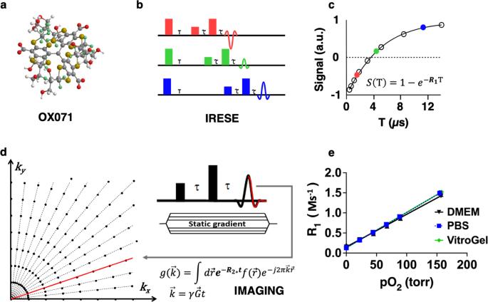
The use of oxygen by cells is an essential aspect of cell metabolism and a reliable indicator of viable and functional cells. Viable and functional cells are essential for optimizing the therapeutic dose for cell therapy, tissue engineering, drug development, and many other biological processes and products. However, currently, there is no method to assess the cell metabolic activity nondestructively in 3D space and longitudinally as cells proliferate, metabolize, differentiate, or die. Here, we report partial pressure oxygen (pO2) mapping of live cells as a reliable indicator of viable and metabolically active cells. For pO2 imaging, we utilized trityl OX071-based pulse electron paramagnetic resonance oxygen imaging (EPROI), in combination with a 25 mT EPROI instrument, JIVA-25™, that provides 3D oxygen maps in tissues with high spatial and temporal resolution. To perform oxygen imaging in an environment-controlled apparatus using a standard biological lab consumable, that is, a multi-well plate, we developed a novel multi-well-plate incubator-resonator (MWIR) system that could accommodate 3 strips from a 96-well strip-well plate and image the middle 12 wells noninvasively and simultaneously. The MWIR system was able to keep a controlled environment (temperature at 37 °C, relative humidity between 70% - 100%, and a controlled gas-flow environment) during oxygen imaging and could keep cells alive for up to 24 h of measurement, providing a rare previously unseen longitudinal perspective of 3D cell metabolic activities. The robustness of MWIR was tested using an adherent cell line (HEK-293 cells), a nonadherent cell line (Jurkat cells), a cell-biomaterial construct (Jurkat cells seeded in a hydrogel), and a negative control (dead HEK-293 cells). Using MWIR, we demonstrate that EPROI is a versatile and robust method that can be utilized to observe the cell metabolic activity nondestructively, longitudinally, and in 3D. For the first time, we demonstrated that oxygen concentration in a multi-well plate seeded with live cells is inversely proportional to the cell seeding density, even if the cells are exposed to incubator-like gas conditions (95% air and 5% CO2). Additionally, for the first time, we also demonstrate 3D, longitudinal oxygen imaging can be used to assess cells seeded in a hydrogel scaffold. These results demonstrate nondestructive, longitudinal 3D assessment of metabolic activities of cells using EPROI during 2D planar culture and during culture in a 3D scaffold system. The MWIR and EPROI approach may be useful for characterizing cell therapies, tissue engineered medical products and other advanced therapeutics.
Abstract Image

利用脉冲电子顺磁共振成像技术对多孔板中的细胞进行无损、纵向、三维氧气成像
细胞对氧气的利用是细胞新陈代谢的一个重要方面,也是细胞存活和功能的一个可靠指标。具有活力和功能的细胞对于优化细胞疗法、组织工程、药物开发以及许多其他生物过程和产品的治疗剂量至关重要。然而,目前还没有一种方法能在三维空间中纵向无损地评估细胞增殖、代谢、分化或死亡过程中的细胞代谢活动。在此,我们报告了活细胞的氧分压(pO2)图谱,它是细胞存活和代谢活跃的可靠指标。为了进行 pO2 成像,我们使用了基于三苯甲基 OX071 的脉冲电子顺磁共振氧成像(EPROI),并结合 25 mT EPROI 仪器 JIVA-25™,该仪器可提供高空间和时间分辨率的组织三维氧图。为了在环境可控的仪器中使用标准生物实验室耗材(即多孔板)进行氧成像,我们开发了一种新型多孔板培养箱-共振器(MWIR)系统,该系统可容纳 96 孔条形孔板中的 3 个条形孔,并对中间的 12 个孔同时进行无创成像。在氧气成像期间,MWIR 系统能够保持受控环境(温度为 37 °C,相对湿度在 70% - 100% 之间,以及受控的气体流动环境),并能使细胞存活长达 24 小时的测量,从而提供了以前从未见过的三维细胞代谢活动纵向视角。我们使用粘附细胞系(HEK-293 细胞)、非粘附细胞系(Jurkat 细胞)、细胞-生物材料构建体(Jurkat 细胞播种在水凝胶中)和阴性对照(死亡的 HEK-293 细胞)测试了 MWIR 的稳健性。通过使用 MWIR,我们证明了 EPROI 是一种多功能、稳健的方法,可用于无损、纵向和三维观察细胞代谢活动。我们首次证明,即使细胞暴露在类似培养箱的气体条件(95% 空气和 5% CO2)下,播种有活细胞的多孔板中的氧气浓度与细胞播种密度成反比。此外,我们还首次证明三维纵向氧气成像可用于评估水凝胶支架中的细胞播种情况。这些结果表明,在二维平面培养和三维支架系统培养过程中,使用 EPROI 可以对细胞的代谢活动进行无损、纵向的三维评估。MWIR 和 EPROI 方法可用于表征细胞疗法、组织工程医疗产品和其他先进疗法。
本文章由计算机程序翻译,如有差异,请以英文原文为准。
求助全文
约1分钟内获得全文 求助全文
来源期刊
自引率
0.00%
发文量
0
×
引用
GB/T 7714-2015
复制
MLA
复制
APA
复制
导出至
BibTeX EndNote RefMan NoteFirst NoteExpress
×
提示
您的信息不完整,为了账户安全,请先补充。
现在去补充
×
提示
您因"违规操作"
具体请查看互助需知
我知道了
×
提示
确定
请完成安全验证×
copy
已复制链接
快去分享给好友吧!
我知道了
右上角分享
点击右上角分享
0
联系我们:info@booksci.cn Book学术提供免费学术资源搜索服务,方便国内外学者检索中英文文献。致力于提供最便捷和优质的服务体验。 Copyright © 2023 布克学术 All rights reserved.
京ICP备2023020795号-1
ghs 京公网安备 11010802042870号
Book学术文献互助
Book学术文献互助群
群 号:481959085
Book学术官方微信