Saede Atarbashi-Moghadam, Ali Lotfi, Parsa Eftekhari-Moghadam
{"title":"Oral Granular Cell Tumor: A Case Report with Emphasis on Pseudoepitheliomatous Hyperplasia in Oral Lesions.","authors":"Saede Atarbashi-Moghadam, Ali Lotfi, Parsa Eftekhari-Moghadam","doi":"10.30476/dentjods.2023.98784.2108","DOIUrl":null,"url":null,"abstract":"<p><p>A granular cell tumor (GCT) is an unusual benign mesenchymal neoplasm with Schwann cells origin. The most common site is the dorsum of the tongue. It has a striking tendency to occur in females and is more frequent in adult patients. GCT typically shows an asymptomatic, slow-growing, single nodule. Histopathologically, it reveals a proliferation of polygonal cells with granular cytoplasm penetrating the adjacent muscles. In some cases, the overlying epithelium demonstrates pseudoepitheliomatous hyperplasia (PEH), which can complicate its precise diagnosis and may mimic squamous cell carcinoma (SCC). This paper presents a 58-year-old woman with a chief complaint of painless mass on the dorsal of the tongue for two years. The lesion was pink and circumscribed with firm consistency measuring 1×1cm. The surface of the lesion was intact. Microscopic examination demonstrated unencapsulated sheets of large, polygonal cells with abundant eosinophilic, granular cytoplasm, and vesicular nuclei. The overlying epithelium showed florid PEH and keratin pearl formation. S100 protein was positive diffusely. The diagnosis of oral GCT was made. Though GCT is a non-aggressive lesion, it may be confused with SCC due to florid PEH and keratin pearl formation. Although PEH is a neglected topic among oral pathologists, it is of great importance in the field of research. Diagnosis can sometimes be problematic because they mimic other lesions. The pathogenesis of PEH is still uncertain. Therefore, familiarity with these characteristics and determining the cause of the PEH leads to correct treatment. This article intends to raise the insight of oral pathologists about PEH in oral lesions.</p>","PeriodicalId":73702,"journal":{"name":"Journal of dentistry (Shiraz, Iran)","volume":"25 1","pages":"91-94"},"PeriodicalIF":0.0000,"publicationDate":"2024-03-01","publicationTypes":"Journal Article","fieldsOfStudy":null,"isOpenAccess":false,"openAccessPdf":"https://www.ncbi.nlm.nih.gov/pmc/articles/PMC10963868/pdf/","citationCount":"0","resultStr":null,"platform":"Semanticscholar","paperid":null,"PeriodicalName":"Journal of dentistry (Shiraz, Iran)","FirstCategoryId":"1085","ListUrlMain":"https://doi.org/10.30476/dentjods.2023.98784.2108","RegionNum":0,"RegionCategory":null,"ArticlePicture":[],"TitleCN":null,"AbstractTextCN":null,"PMCID":null,"EPubDate":"","PubModel":"","JCR":"","JCRName":"","Score":null,"Total":0}
引用次数: 0
Abstract
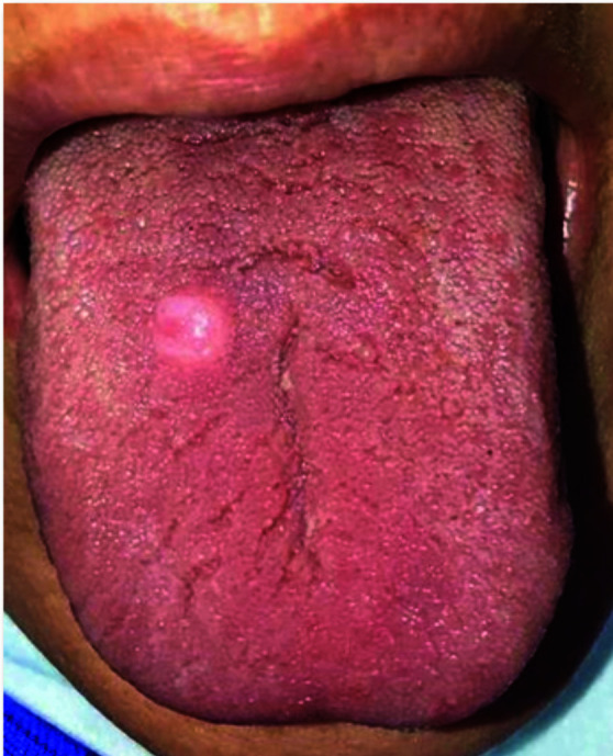
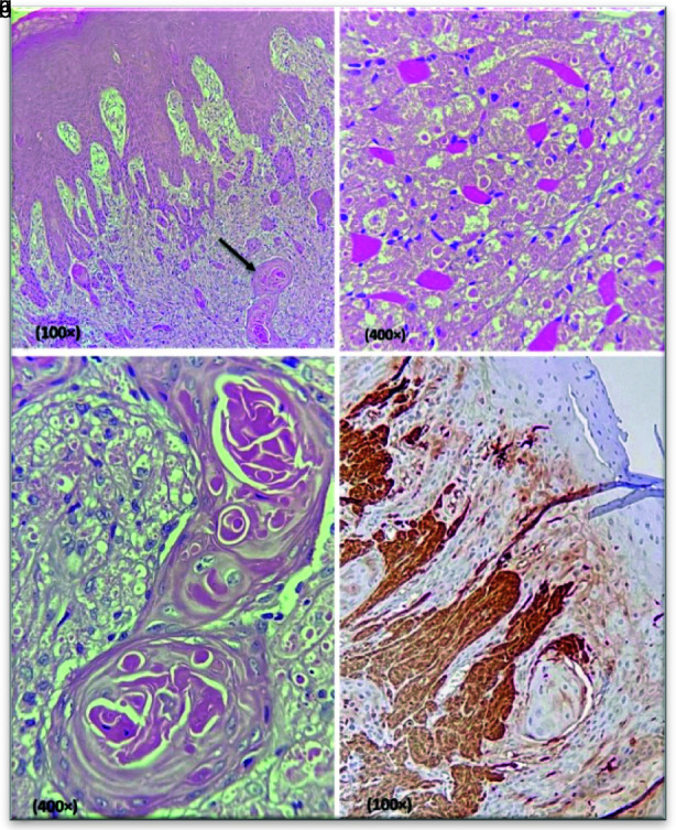
A granular cell tumor (GCT) is an unusual benign mesenchymal neoplasm with Schwann cells origin. The most common site is the dorsum of the tongue. It has a striking tendency to occur in females and is more frequent in adult patients. GCT typically shows an asymptomatic, slow-growing, single nodule. Histopathologically, it reveals a proliferation of polygonal cells with granular cytoplasm penetrating the adjacent muscles. In some cases, the overlying epithelium demonstrates pseudoepitheliomatous hyperplasia (PEH), which can complicate its precise diagnosis and may mimic squamous cell carcinoma (SCC). This paper presents a 58-year-old woman with a chief complaint of painless mass on the dorsal of the tongue for two years. The lesion was pink and circumscribed with firm consistency measuring 1×1cm. The surface of the lesion was intact. Microscopic examination demonstrated unencapsulated sheets of large, polygonal cells with abundant eosinophilic, granular cytoplasm, and vesicular nuclei. The overlying epithelium showed florid PEH and keratin pearl formation. S100 protein was positive diffusely. The diagnosis of oral GCT was made. Though GCT is a non-aggressive lesion, it may be confused with SCC due to florid PEH and keratin pearl formation. Although PEH is a neglected topic among oral pathologists, it is of great importance in the field of research. Diagnosis can sometimes be problematic because they mimic other lesions. The pathogenesis of PEH is still uncertain. Therefore, familiarity with these characteristics and determining the cause of the PEH leads to correct treatment. This article intends to raise the insight of oral pathologists about PEH in oral lesions.

 求助内容:
求助内容: 应助结果提醒方式:


