{"title":"Histopathologic Evaluation of Periapical Radiolucencies Clinico-Radiographically Diagnosed as Endodontic Lesions: A Retrospective Analysis.","authors":"Saede Atarbashi-Moghadam, Mehrdad Azar, Shaghayegheh Dowdani","doi":"10.30476/dentjods.2023.96819.1967","DOIUrl":null,"url":null,"abstract":"<p><strong>Statement of the problem: </strong>Periapical cyst and granuloma are inflammatory endodontic lesions. Periapical granuloma usually heals spontaneously after endodontic treatment; however, periapical cyst mostly needs to be removed via surgical approaches. Although some clinicians believe that microscopic examination of periapical lesions is unnecessary, it is proved that some of them has non-endodontic nature that need critical consideration.</p><p><strong>Purpose: </strong>The purpose of this study was to assess the disagreement between clinico-radiographic and microscopic diagnosis of periapical cysts and granulomas in a major center of oral pathology service in Iran.</p><p><strong>Materials and method: </strong>In this retrospective, descriptive cross-sectional study, the archives of the oral and maxillofacial pathology department of Shahid Beheshti University of Medical Sciences served as the source of the material during an 18-year-period for this retrospective, descriptive cross-sectional study. The reports of all patients whose initial clinical diagnosis was a periapical cyst/granuloma were extracted.</p><p><strong>Results: </strong> In the present study, 474 cases were diagnosed with a periapical cyst/granuloma clinico-radiographically, of which 61 cases (12.86%) received a microscopic diagnosis of a non-endodontic pathology. The most frequent lesion was odontogenic keratocyst (n= 12, 19.67%) followed by infected odontogenic cyst (n= 12, 19.67%). About 21.31% of diagnoses were non-cystic lesions and 4.9% were malignancies. The most odontogenic tumors that were diagnosed as periapical cyst/granuloma in clinico-radiography were the ameloblastoma variants (n= 4, 6.55%).</p><p><strong>Conclusion: </strong> A wide variety of microscopic diagnoses, including aggressive lesions such as ameloblastoma, as well as other malignant lesions was noted in this study. These misdiagnoses can lead to an inappropriate treatment plan. It is important to microscopically examine all lesions removed from the jaw.</p>","PeriodicalId":73702,"journal":{"name":"Journal of dentistry (Shiraz, Iran)","volume":"25 1","pages":"39-44"},"PeriodicalIF":0.0000,"publicationDate":"2024-03-01","publicationTypes":"Journal Article","fieldsOfStudy":null,"isOpenAccess":false,"openAccessPdf":"https://www.ncbi.nlm.nih.gov/pmc/articles/PMC10963870/pdf/","citationCount":"0","resultStr":null,"platform":"Semanticscholar","paperid":null,"PeriodicalName":"Journal of dentistry (Shiraz, Iran)","FirstCategoryId":"1085","ListUrlMain":"https://doi.org/10.30476/dentjods.2023.96819.1967","RegionNum":0,"RegionCategory":null,"ArticlePicture":[],"TitleCN":null,"AbstractTextCN":null,"PMCID":null,"EPubDate":"","PubModel":"","JCR":"","JCRName":"","Score":null,"Total":0}
引用次数: 0
Abstract
Statement of the problem: Periapical cyst and granuloma are inflammatory endodontic lesions. Periapical granuloma usually heals spontaneously after endodontic treatment; however, periapical cyst mostly needs to be removed via surgical approaches. Although some clinicians believe that microscopic examination of periapical lesions is unnecessary, it is proved that some of them has non-endodontic nature that need critical consideration.
Purpose: The purpose of this study was to assess the disagreement between clinico-radiographic and microscopic diagnosis of periapical cysts and granulomas in a major center of oral pathology service in Iran.
Materials and method: In this retrospective, descriptive cross-sectional study, the archives of the oral and maxillofacial pathology department of Shahid Beheshti University of Medical Sciences served as the source of the material during an 18-year-period for this retrospective, descriptive cross-sectional study. The reports of all patients whose initial clinical diagnosis was a periapical cyst/granuloma were extracted.
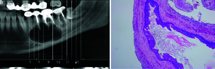
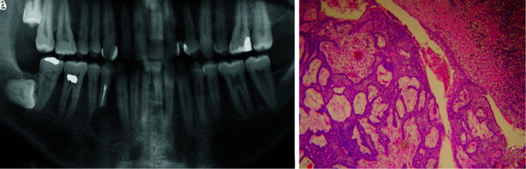
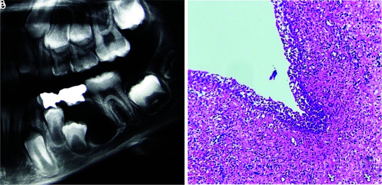
Results: In the present study, 474 cases were diagnosed with a periapical cyst/granuloma clinico-radiographically, of which 61 cases (12.86%) received a microscopic diagnosis of a non-endodontic pathology. The most frequent lesion was odontogenic keratocyst (n= 12, 19.67%) followed by infected odontogenic cyst (n= 12, 19.67%). About 21.31% of diagnoses were non-cystic lesions and 4.9% were malignancies. The most odontogenic tumors that were diagnosed as periapical cyst/granuloma in clinico-radiography were the ameloblastoma variants (n= 4, 6.55%).
Conclusion: A wide variety of microscopic diagnoses, including aggressive lesions such as ameloblastoma, as well as other malignant lesions was noted in this study. These misdiagnoses can lead to an inappropriate treatment plan. It is important to microscopically examine all lesions removed from the jaw.

 求助内容:
求助内容: 应助结果提醒方式:


