Bhargav D Sanketi, Madhav Mantri, Liqing Huang, Mohammad A Tavallaei, Shing Hu, Michael F Z Wang, Iwijn De Vlaminck, Natasza A Kurpios
{"title":"Villus myofibroblasts are developmental and adult progenitors of mammalian gut lymphatic musculature.","authors":"Bhargav D Sanketi, Madhav Mantri, Liqing Huang, Mohammad A Tavallaei, Shing Hu, Michael F Z Wang, Iwijn De Vlaminck, Natasza A Kurpios","doi":"10.1016/j.devcel.2024.03.005","DOIUrl":null,"url":null,"abstract":"<p><p>Inside the finger-like intestinal projections called villi, strands of smooth muscle cells contract to propel absorbed dietary fats through the adjacent lymphatic capillary, the lacteal, sending fats into the systemic blood circulation for energy production. Despite this vital function, mechanisms of formation, assembly alongside lacteals, and maintenance of villus smooth muscle are unknown. By combining single-cell RNA sequencing and quantitative lineage tracing of the mouse intestine, we identified a local hierarchy of subepithelial fibroblast progenitors that differentiate into mature smooth muscle fibers via intermediate contractile myofibroblasts. This continuum persists as the major mechanism for villus musculature renewal throughout adult life. The NOTCH3-DLL4 signaling axis governs the assembly of smooth muscle fibers alongside their adjacent lacteals and is required for fat absorption. Our studies identify the ontogeny and maintenance of a poorly defined class of intestinal smooth muscle, with implications for accelerated repair and recovery of digestive function following injury.</p>","PeriodicalId":11157,"journal":{"name":"Developmental cell","volume":null,"pages":null},"PeriodicalIF":10.7000,"publicationDate":"2024-05-06","publicationTypes":"Journal Article","fieldsOfStudy":null,"isOpenAccess":false,"openAccessPdf":"https://www.ncbi.nlm.nih.gov/pmc/articles/PMC11078612/pdf/","citationCount":"0","resultStr":null,"platform":"Semanticscholar","paperid":null,"PeriodicalName":"Developmental cell","FirstCategoryId":"99","ListUrlMain":"https://doi.org/10.1016/j.devcel.2024.03.005","RegionNum":1,"RegionCategory":"生物学","ArticlePicture":[],"TitleCN":null,"AbstractTextCN":null,"PMCID":null,"EPubDate":"2024/3/26 0:00:00","PubModel":"Epub","JCR":"Q1","JCRName":"CELL BIOLOGY","Score":null,"Total":0}
引用次数: 0
Abstract
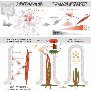
Inside the finger-like intestinal projections called villi, strands of smooth muscle cells contract to propel absorbed dietary fats through the adjacent lymphatic capillary, the lacteal, sending fats into the systemic blood circulation for energy production. Despite this vital function, mechanisms of formation, assembly alongside lacteals, and maintenance of villus smooth muscle are unknown. By combining single-cell RNA sequencing and quantitative lineage tracing of the mouse intestine, we identified a local hierarchy of subepithelial fibroblast progenitors that differentiate into mature smooth muscle fibers via intermediate contractile myofibroblasts. This continuum persists as the major mechanism for villus musculature renewal throughout adult life. The NOTCH3-DLL4 signaling axis governs the assembly of smooth muscle fibers alongside their adjacent lacteals and is required for fat absorption. Our studies identify the ontogeny and maintenance of a poorly defined class of intestinal smooth muscle, with implications for accelerated repair and recovery of digestive function following injury.
期刊介绍:
Developmental Cell, established in 2001, is a comprehensive journal that explores a wide range of topics in cell and developmental biology. Our publication encompasses work across various disciplines within biology, with a particular emphasis on investigating the intersections between cell biology, developmental biology, and other related fields. Our primary objective is to present research conducted through a cell biological perspective, addressing the essential mechanisms governing cell function, cellular interactions, and responses to the environment. Moreover, we focus on understanding the collective behavior of cells, culminating in the formation of tissues, organs, and whole organisms, while also investigating the consequences of any malfunctions in these intricate processes.
 求助内容:
求助内容: 应助结果提醒方式:


