{"title":"In vitro comparison of accuracy between conventional and digital impression using elastomeric materials and two intra-oral scanning devices","authors":"Eirini Palantza DDS, MSc, Nikitas Sykaras DDS, PhD, Panagiotis Zoidis DDS, MSc, PhD, Stefanos Kourtis DDS, Dr. Odont","doi":"10.1111/jerd.13227","DOIUrl":null,"url":null,"abstract":"<div>\n \n \n <section>\n \n <h3> Aim</h3>\n \n <p>The aim of this study was to compare the accuracy of full-arch conventional implant impressions using two different materials (A-silicone and polyether) to full-arch digital implant impressions produced from two intraoral scanning devices.</p>\n </section>\n \n <section>\n \n <h3> Materials and Methods</h3>\n \n <p>A master model was fabricated representing an edentulous mandible with four implants with internal connection placed at the sites of canines and first molars. The anterior implants were parallel to the residual ridge, while the two posterior implants had an angulation of 15° to the distal and 15° to the lingual respectively. The conventional technique was performed with open-tray of non-splinted impression copings. Two different impression materials were used, A-silicone and polyether at monophase medium body consistencies. The digital impressions were obtained with the use of two different intraoral scanners, after the connection of scan bodies. A total of 10 impressions were produced for each of the four experimental groups. The conventional models as well as the master model were digitized using a high-resolution laboratory scanner. The STL files of the models and of the intraoral impressions were imported in a powerful superimposition software, for the conduction of measurements in pairs of files. The software calculated the 3D deviations, as well as the linear and angular displacements among scan bodies at the digital files. For “trueness” measurements every STL file of each experimental group was superimposed to the digital master model, while for “precision” measurements all STL files of each experimental group were superimposed to each other.</p>\n </section>\n \n <section>\n \n <h3> Results and Conclusions</h3>\n \n <div>\n <ul>\n \n <li>The accuracy of full arch mandibular implant impressions was influenced both by the impression technique used (conventional vs. digital) and the impression material used (A-silicone vs. polyether) or the intraoral scanner used (Trios vs. Heron).</li>\n \n <li>In terms of “trueness,” A-silicone showed the highest impression accuracy with the lowest deviation values, followed by polyether and Trios, but the differences between the three groups were in the majority not statistically significant. Heron showed statistically lower accuracy results in all measurements compared to the other groups.</li>\n \n <li>In terms of “precision”, conventional impressions with the use of A-Silicone or polyether were statistically significantly superior to digital impressions with either scanner. A-Silicone and polyether showed no statistically significant difference between them.</li>\n </ul>\n </div>\n </section>\n </div>","PeriodicalId":15988,"journal":{"name":"Journal of Esthetic and Restorative Dentistry","volume":null,"pages":null},"PeriodicalIF":3.2000,"publicationDate":"2024-03-27","publicationTypes":"Journal Article","fieldsOfStudy":null,"isOpenAccess":false,"openAccessPdf":"https://onlinelibrary.wiley.com/doi/epdf/10.1111/jerd.13227","citationCount":"0","resultStr":null,"platform":"Semanticscholar","paperid":null,"PeriodicalName":"Journal of Esthetic and Restorative Dentistry","FirstCategoryId":"3","ListUrlMain":"https://onlinelibrary.wiley.com/doi/10.1111/jerd.13227","RegionNum":3,"RegionCategory":"医学","ArticlePicture":[],"TitleCN":null,"AbstractTextCN":null,"PMCID":null,"EPubDate":"","PubModel":"","JCR":"Q1","JCRName":"DENTISTRY, ORAL SURGERY & MEDICINE","Score":null,"Total":0}
引用次数: 0
Abstract
Aim
The aim of this study was to compare the accuracy of full-arch conventional implant impressions using two different materials (A-silicone and polyether) to full-arch digital implant impressions produced from two intraoral scanning devices.
Materials and Methods
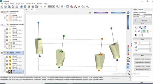
A master model was fabricated representing an edentulous mandible with four implants with internal connection placed at the sites of canines and first molars. The anterior implants were parallel to the residual ridge, while the two posterior implants had an angulation of 15° to the distal and 15° to the lingual respectively. The conventional technique was performed with open-tray of non-splinted impression copings. Two different impression materials were used, A-silicone and polyether at monophase medium body consistencies. The digital impressions were obtained with the use of two different intraoral scanners, after the connection of scan bodies. A total of 10 impressions were produced for each of the four experimental groups. The conventional models as well as the master model were digitized using a high-resolution laboratory scanner. The STL files of the models and of the intraoral impressions were imported in a powerful superimposition software, for the conduction of measurements in pairs of files. The software calculated the 3D deviations, as well as the linear and angular displacements among scan bodies at the digital files. For “trueness” measurements every STL file of each experimental group was superimposed to the digital master model, while for “precision” measurements all STL files of each experimental group were superimposed to each other.
Results and Conclusions
The accuracy of full arch mandibular implant impressions was influenced both by the impression technique used (conventional vs. digital) and the impression material used (A-silicone vs. polyether) or the intraoral scanner used (Trios vs. Heron).
In terms of “trueness,” A-silicone showed the highest impression accuracy with the lowest deviation values, followed by polyether and Trios, but the differences between the three groups were in the majority not statistically significant. Heron showed statistically lower accuracy results in all measurements compared to the other groups.
In terms of “precision”, conventional impressions with the use of A-Silicone or polyether were statistically significantly superior to digital impressions with either scanner. A-Silicone and polyether showed no statistically significant difference between them.
期刊介绍:
The Journal of Esthetic and Restorative Dentistry (JERD) is the longest standing peer-reviewed journal devoted solely to advancing the knowledge and practice of esthetic dentistry. Its goal is to provide the very latest evidence-based information in the realm of contemporary interdisciplinary esthetic dentistry through high quality clinical papers, sound research reports and educational features.
The range of topics covered in the journal includes:
- Interdisciplinary esthetic concepts
- Implants
- Conservative adhesive restorations
- Tooth Whitening
- Prosthodontic materials and techniques
- Dental materials
- Orthodontic, periodontal and endodontic esthetics
- Esthetics related research
- Innovations in esthetics