Free flap reconstruction of elbow soft tissue defects: Lessons learned from 15 years of experience

IF 1.5 3区 医学 Q3 SURGERY
Microsurgery Pub Date : 2024-03-26 DOI:10.1002/micr.31163
J. Reed McGraw BS, Reena S. Sulkar MBA, Corey M. Bascone MD, MBA, Sammy Othman MD, Jaclyn T. Mauch MD, MBE, Hani I. Naga MD, L. Scott Levin MD, Stephen J. Kovach III MD
{"title":"Free flap reconstruction of elbow soft tissue defects: Lessons learned from 15 years of experience","authors":"J. Reed McGraw BS,&nbsp;Reena S. Sulkar MBA,&nbsp;Corey M. Bascone MD, MBA,&nbsp;Sammy Othman MD,&nbsp;Jaclyn T. Mauch MD, MBE,&nbsp;Hani I. Naga MD,&nbsp;L. Scott Levin MD,&nbsp;Stephen J. Kovach III MD","doi":"10.1002/micr.31163","DOIUrl":null,"url":null,"abstract":"<div>\n \n \n <section>\n \n <h3> Background</h3>\n \n <p>The elbow is a complex joint that is vital for proper function of the upper extremity. Reconstruction of soft tissue defects over the joint space remains challenging, and outcomes following free tissue transfer remain underreported in the literature. The purpose of this analysis was to evaluate the rate of limb salvage, joint function, and clinical complications following microvascular free flap coverage of the elbow.</p>\n </section>\n \n <section>\n \n <h3> Methods</h3>\n \n <p>This retrospective case series utilized surgical logs of the senior authors (Stephen J Kovach and L Scott Levin) to identify patients who underwent microvascular free flap elbow reconstruction between January 2007 and December 2021. Patient demographics and medical history were collected from the medical chart. Operative notes were reviewed to determine the type of flap procedure performed. The achievement of definitive soft tissue coverage, joint function, and limb salvage status at 1 year was determined from postoperative visit notes.</p>\n </section>\n \n <section>\n \n <h3> Results</h3>\n \n <p>Twenty-one patients (14 male, 7 female, median age 43) underwent free tissue transfer for coverage of soft tissue defects of the elbow. The most common indication for free tissue transfer was traumatic elbow fracture with soft tissue loss (<i>n</i> = 12, [57%]). Among the 21 free flaps performed, 71% (<i>n</i> = 15) were anterolateral thigh flaps, 14% (<i>n</i> = 3) were latissimus dorsi flaps, and 5% (<i>n</i> = 1) were transverse rectus abdominis flaps. The mean flap size was 107.5 cm<sup>2</sup>. Flap success was 100% (<i>n</i> = 21). The following postoperative wound complications were reported: surgical site infection (<i>n</i> = 1, [5%]); partial dehiscence (<i>n</i> = 5, [24%]); seroma (<i>n</i> = 2, [10%]); donor-site hematoma (<i>n</i> = 1, [5%]); and delayed wound healing (<i>n</i> = 5, [24%]). At 1 year, all 21 patients achieved limb salvage and definitive soft tissue coverage. Of the 17 patients with functional data available, 47% (<i>n</i> = 8) had regained at least 120 degrees of elbow flexion/extension. All patients had greater than 1 year of follow-up.</p>\n </section>\n \n <section>\n \n <h3> Conclusion</h3>\n \n <p>Microvascular free flap reconstruction is a safe and effective method of providing definitive soft tissue coverage of elbow defects, as evidenced by high rates of limb salvage and functional recovery following reconstruction.</p>\n </section>\n </div>","PeriodicalId":18600,"journal":{"name":"Microsurgery","volume":null,"pages":null},"PeriodicalIF":1.5000,"publicationDate":"2024-03-26","publicationTypes":"Journal Article","fieldsOfStudy":null,"isOpenAccess":false,"openAccessPdf":"https://onlinelibrary.wiley.com/doi/epdf/10.1002/micr.31163","citationCount":"0","resultStr":null,"platform":"Semanticscholar","paperid":null,"PeriodicalName":"Microsurgery","FirstCategoryId":"3","ListUrlMain":"https://onlinelibrary.wiley.com/doi/10.1002/micr.31163","RegionNum":3,"RegionCategory":"医学","ArticlePicture":[],"TitleCN":null,"AbstractTextCN":null,"PMCID":null,"EPubDate":"","PubModel":"","JCR":"Q3","JCRName":"SURGERY","Score":null,"Total":0}
引用次数: 0

Abstract

Background

The elbow is a complex joint that is vital for proper function of the upper extremity. Reconstruction of soft tissue defects over the joint space remains challenging, and outcomes following free tissue transfer remain underreported in the literature. The purpose of this analysis was to evaluate the rate of limb salvage, joint function, and clinical complications following microvascular free flap coverage of the elbow.

Methods

This retrospective case series utilized surgical logs of the senior authors (Stephen J Kovach and L Scott Levin) to identify patients who underwent microvascular free flap elbow reconstruction between January 2007 and December 2021. Patient demographics and medical history were collected from the medical chart. Operative notes were reviewed to determine the type of flap procedure performed. The achievement of definitive soft tissue coverage, joint function, and limb salvage status at 1 year was determined from postoperative visit notes.

Results

Twenty-one patients (14 male, 7 female, median age 43) underwent free tissue transfer for coverage of soft tissue defects of the elbow. The most common indication for free tissue transfer was traumatic elbow fracture with soft tissue loss (n = 12, [57%]). Among the 21 free flaps performed, 71% (n = 15) were anterolateral thigh flaps, 14% (n = 3) were latissimus dorsi flaps, and 5% (n = 1) were transverse rectus abdominis flaps. The mean flap size was 107.5 cm2. Flap success was 100% (n = 21). The following postoperative wound complications were reported: surgical site infection (n = 1, [5%]); partial dehiscence (n = 5, [24%]); seroma (n = 2, [10%]); donor-site hematoma (n = 1, [5%]); and delayed wound healing (n = 5, [24%]). At 1 year, all 21 patients achieved limb salvage and definitive soft tissue coverage. Of the 17 patients with functional data available, 47% (n = 8) had regained at least 120 degrees of elbow flexion/extension. All patients had greater than 1 year of follow-up.

Conclusion

Microvascular free flap reconstruction is a safe and effective method of providing definitive soft tissue coverage of elbow defects, as evidenced by high rates of limb salvage and functional recovery following reconstruction.
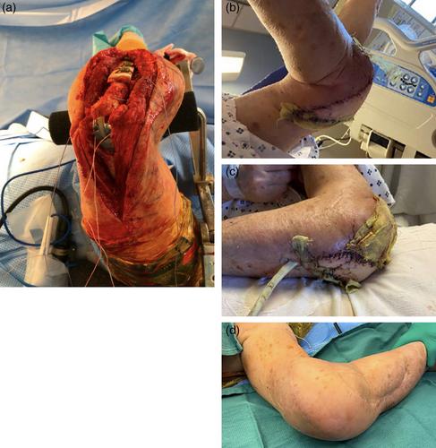
Abstract Image

肘部软组织缺损的游离皮瓣重建:从 15 年的经验中汲取教训。
背景:肘部是一个复杂的关节,对上肢的正常功能至关重要。关节间隙软组织缺损的重建仍具有挑战性,文献中对游离组织转移后的结果报道不足。本分析旨在评估肘部微血管游离皮瓣覆盖后的肢体挽救率、关节功能和临床并发症:这项回顾性病例系列研究利用资深作者(Stephen J Kovach 和 L Scott Levin)的手术日志确定了在 2007 年 1 月至 2021 年 12 月期间接受微血管游离皮瓣肘关节重建术的患者。从病历中收集了患者的人口统计学特征和病史。审查手术记录以确定所实施的皮瓣手术类型。根据术后访视记录确定1年后软组织覆盖、关节功能和肢体挽回情况:21名患者(14名男性,7名女性,中位年龄43岁)接受了游离组织转移术,以覆盖肘部软组织缺损。最常见的游离组织转移适应症是外伤性肘部骨折伴软组织缺损(12 例,[57%])。在21个游离皮瓣中,71%(15个)为大腿前外侧皮瓣,14%(3个)为背阔肌皮瓣,5%(1个)为腹横肌皮瓣。皮瓣的平均大小为 107.5 平方厘米。皮瓣成功率为 100%(n = 21)。术后伤口并发症如下:手术部位感染(1例,[5%]);部分开裂(5例,[24%]);血清肿(2例,[10%]);供体部位血肿(1例,[5%]);伤口延迟愈合(5例,[24%])。1 年后,所有 21 名患者都实现了肢体挽救和明确的软组织覆盖。在17名有功能数据的患者中,47%(8人)的肘关节屈伸功能恢复了至少120度。所有患者的随访时间均超过1年:微血管游离皮瓣重建是一种安全有效的方法,可为肘部缺损提供明确的软组织覆盖,重建后的肢体挽救率和功能恢复率都很高。
本文章由计算机程序翻译,如有差异,请以英文原文为准。
求助全文
约1分钟内获得全文 求助全文
来源期刊
Microsurgery
Microsurgery 医学-外科
CiteScore
3.80
自引率
19.00%
发文量
128
审稿时长
4-8 weeks
期刊介绍: Microsurgery is an international and interdisciplinary publication of original contributions concerning surgery under microscopic magnification. Microsurgery publishes clinical studies, research papers, invited articles, relevant reviews, and other scholarly works from all related fields including orthopaedic surgery, otolaryngology, pediatric surgery, plastic surgery, urology, and vascular surgery.
×
引用
GB/T 7714-2015
复制
MLA
复制
APA
复制
导出至
BibTeX EndNote RefMan NoteFirst NoteExpress
×
提示
您的信息不完整,为了账户安全,请先补充。
现在去补充
×
提示
您因"违规操作"
具体请查看互助需知
我知道了
×
提示
确定
请完成安全验证×
copy
已复制链接
快去分享给好友吧!
我知道了
右上角分享
点击右上角分享
0
联系我们:info@booksci.cn Book学术提供免费学术资源搜索服务,方便国内外学者检索中英文文献。致力于提供最便捷和优质的服务体验。 Copyright © 2023 布克学术 All rights reserved.
京ICP备2023020795号-1
ghs 京公网安备 11010802042870号
Book学术文献互助
Book学术文献互助群
群 号:481959085
Book学术官方微信