Three-dimensional wall-thickness distributions of unruptured intracranial aneurysms characterized by micro-computed tomography
Abstract
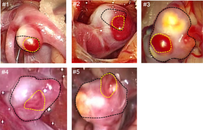
Aneurysmal rupture is associated with wall thinning, but the mechanism is poorly understood. This study aimed to characterize the three-dimensional wall-thickness distributions of unruptured intracranial aneurysms. Five aneurysmal tissues were investigated using micro-computed tomography. First, the wall thickness was related to the aneurysmal wall appearances during surgery. The median wall thicknesses of the translucent and non-translucent walls were 50.56 and 155.93 µm, respectively (p < 0.05) with significant variation in the non-translucent wall thicknesses (p < 0.05). The three-dimensional observations characterized the spatial variation of wall thicknesses. Thin walls showed a uniform thickness profile ranging from 10 to 40 µm, whereas thick walls presented a peaked thickness profile ranging from 300 to 500 µm. In transition walls, the profile undulated due to the formation of focal thin/thick spots. Overall, the aneurysmal wall thicknesses were strongly site-dependent and spatially varied by 10 to 40 times within individual cases. Aneurysmal walls are exposed to wall stress driven by blood pressure. In theory, the magnitude of wall stress is inversely proportional to wall thickness. Thus, the observed spatial variation of wall thickness may increase the spatial variation of wall stress to a similar extent. The irregular wall thickness may yield stress concentration. The observed thin walls and focal thin spots may be caused by excessive wall stresses at the range of mechanical failure inducing wall injuries, such as microscopic tears, during aneurysmal enlargement. The present results suggested that blood pressure (wall stress) may have a potential of acting as a trigger of aneurysmal wall injury.


 求助内容:
求助内容: 应助结果提醒方式:


