Leonard W.F. Seelen MD, Lieke van den Wildenberg MS, Ayhan Gursan MS, Martijn Froeling PhD, Mark W.J.M. Gosselink MS, Wybe J.M. van der Kemp PhD, Nadia Haj Mohammad MD, PhD, I. Quintus Molenaar MD, PhD, Hjalmar C. van Santvoort MD, PhD, Dennis W.J. Klomp PhD, Jeanine J. Prompers PhD
{"title":"31P MR Spectroscopy in the Pancreas: Repeatability, Comparison With Liver, and Pilot Pancreatic Cancer Data","authors":"Leonard W.F. Seelen MD, Lieke van den Wildenberg MS, Ayhan Gursan MS, Martijn Froeling PhD, Mark W.J.M. Gosselink MS, Wybe J.M. van der Kemp PhD, Nadia Haj Mohammad MD, PhD, I. Quintus Molenaar MD, PhD, Hjalmar C. van Santvoort MD, PhD, Dennis W.J. Klomp PhD, Jeanine J. Prompers PhD","doi":"10.1002/jmri.29326","DOIUrl":null,"url":null,"abstract":"<div>\n \n <section>\n \n <h3> Background</h3>\n \n <p>Non-invasive evaluation of phosphomonoesters (PMEs) and phosphodiesters (PDEs) by 31-phosphorus MR spectroscopy (<sup>31</sup>P MRS) may have potential for early therapy (non-)response assessment in cancer. However, <sup>31</sup>P MRS has not yet been applied to investigate the human pancreas <i>in vivo</i>.</p>\n </section>\n \n <section>\n \n <h3> Purpose</h3>\n \n <p>To assess the technical feasibility and repeatability of <sup>31</sup>P MR spectroscopic imaging (MRSI) of the pancreas, compare <sup>31</sup>P metabolite levels between pancreas and liver, and determine the feasibility of <sup>31</sup>P MRSI in pancreatic cancer.</p>\n </section>\n \n <section>\n \n <h3> Study Type</h3>\n \n <p>Prospective cohort study.</p>\n </section>\n \n <section>\n \n <h3> Population</h3>\n \n <p>10 healthy subjects (age 34 ± 12 years, four females) and one patient (73-year-old female) with pancreatic ductal adenocarcinoma.</p>\n </section>\n \n <section>\n \n <h3> Field Strength/Sequence</h3>\n \n <p>7-T, <sup>31</sup>P FID-MRSI, <sup>1</sup>H gradient-echo MRI.</p>\n </section>\n \n <section>\n \n <h3> Assessment</h3>\n \n <p><sup>31</sup>P FID-MRSI of the abdomen (including the pancreas and liver) was performed with a nominal voxel size of 20 mm (isotropic). For repeatability measurements, healthy subjects were scanned twice on the same day. The patient was only scanned once. Test–retest <sup>31</sup>P MRSI data of pancreas and liver voxels (segmented on <sup>1</sup>H MRI) of healthy subjects were quantified by fitting in the time domain and signal amplitudes were normalized to γ-adenosine triphosphate. In addition, the PME/PDE ratio was calculated. Metabolite levels were averaged over all voxels within the pancreas, right liver lobe and left liver lobe, respectively.</p>\n </section>\n \n <section>\n \n <h3> Statistical Tests</h3>\n \n <p>Repeatability of test–retest data from healthy pancreas was assessed by paired <i>t</i>-tests, Bland–Altman analyses, and calculation of the intrasubject coefficients of variation (CoVs). Significant differences between healthy pancreas and right and left liver lobes were assessed with a two-way analysis of variance (ANOVA) for repeated measures. A <i>P</i>-value <0.05 was considered statistically significant.</p>\n </section>\n \n <section>\n \n <h3> Results</h3>\n \n <p>The intrasubject CoVs for PME, PDE, and PME/PDE in healthy pancreas were below 20%. Furthermore, PME and PME/PDE were significantly higher in pancreas compared to liver. In the patient with pancreatic cancer, qualitatively, elevated relative PME signals were observed in comparison with healthy pancreas.</p>\n </section>\n \n <section>\n \n <h3> Data Conclusion</h3>\n \n <p><i>In vivo</i> <sup>31</sup>P MRSI of the human healthy pancreas and in pancreatic cancer may be feasible at 7 T.</p>\n </section>\n \n <section>\n \n <h3> Evidence Level</h3>\n \n <p>3</p>\n </section>\n \n <section>\n \n <h3> Technical Efficacy</h3>\n \n <p>Stage 2</p>\n </section>\n </div>","PeriodicalId":3,"journal":{"name":"ACS Applied Electronic Materials","volume":null,"pages":null},"PeriodicalIF":4.3000,"publicationDate":"2024-03-14","publicationTypes":"Journal Article","fieldsOfStudy":null,"isOpenAccess":false,"openAccessPdf":"https://onlinelibrary.wiley.com/doi/epdf/10.1002/jmri.29326","citationCount":"0","resultStr":null,"platform":"Semanticscholar","paperid":null,"PeriodicalName":"ACS Applied Electronic Materials","FirstCategoryId":"3","ListUrlMain":"https://onlinelibrary.wiley.com/doi/10.1002/jmri.29326","RegionNum":3,"RegionCategory":"材料科学","ArticlePicture":[],"TitleCN":null,"AbstractTextCN":null,"PMCID":null,"EPubDate":"","PubModel":"","JCR":"Q1","JCRName":"ENGINEERING, ELECTRICAL & ELECTRONIC","Score":null,"Total":0}
引用次数: 0
Abstract
Background
Non-invasive evaluation of phosphomonoesters (PMEs) and phosphodiesters (PDEs) by 31-phosphorus MR spectroscopy (31P MRS) may have potential for early therapy (non-)response assessment in cancer. However, 31P MRS has not yet been applied to investigate the human pancreas in vivo.
Purpose
To assess the technical feasibility and repeatability of 31P MR spectroscopic imaging (MRSI) of the pancreas, compare 31P metabolite levels between pancreas and liver, and determine the feasibility of 31P MRSI in pancreatic cancer.
Study Type
Prospective cohort study.
Population
10 healthy subjects (age 34 ± 12 years, four females) and one patient (73-year-old female) with pancreatic ductal adenocarcinoma.
Field Strength/Sequence
7-T, 31P FID-MRSI, 1H gradient-echo MRI.
Assessment
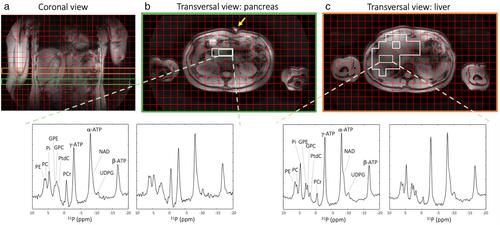
31P FID-MRSI of the abdomen (including the pancreas and liver) was performed with a nominal voxel size of 20 mm (isotropic). For repeatability measurements, healthy subjects were scanned twice on the same day. The patient was only scanned once. Test–retest 31P MRSI data of pancreas and liver voxels (segmented on 1H MRI) of healthy subjects were quantified by fitting in the time domain and signal amplitudes were normalized to γ-adenosine triphosphate. In addition, the PME/PDE ratio was calculated. Metabolite levels were averaged over all voxels within the pancreas, right liver lobe and left liver lobe, respectively.
Statistical Tests
Repeatability of test–retest data from healthy pancreas was assessed by paired t-tests, Bland–Altman analyses, and calculation of the intrasubject coefficients of variation (CoVs). Significant differences between healthy pancreas and right and left liver lobes were assessed with a two-way analysis of variance (ANOVA) for repeated measures. A P-value <0.05 was considered statistically significant.
Results
The intrasubject CoVs for PME, PDE, and PME/PDE in healthy pancreas were below 20%. Furthermore, PME and PME/PDE were significantly higher in pancreas compared to liver. In the patient with pancreatic cancer, qualitatively, elevated relative PME signals were observed in comparison with healthy pancreas.
Data Conclusion
In vivo31P MRSI of the human healthy pancreas and in pancreatic cancer may be feasible at 7 T.