{"title":"Regional differences in the ultrastructure of mucosal macrophages in the rat large intestine.","authors":"Shota Murase, Youhei Mantani, Nobuhiko Ohno, Asaka Shimada, Satoki Nakanishi, Rinako Morishita, Toshifumi Yokoyama, Nobuhiko Hoshi","doi":"10.1007/s00441-024-03883-w","DOIUrl":null,"url":null,"abstract":"<p><p>We previously clarified the histological characteristics of macrophages in the rat small intestine using serial block-face scanning electron microscopy (SBF-SEM). However, the regional differences in the characteristics of macrophages throughout the large intestine remain unknown. Here, we performed a pilot study to explore the regional differences in the ultrastructure of mucosal macrophages in the large intestine by using SBF-SEM analysis. SBF-SEM analysis conducted on the luminal side of the cecum and descending colon revealed macrophages as amorphous cells possessing abundant lysosomes and vacuoles. Macrophages in the cecum exhibited a higher abundance of lysosomes and a lower abundance of vacuoles than those in the descending colon. Macrophages with many intraepithelial cellular processes were observed beneath the intestinal superficial epithelium in the descending colon. Moreover, macrophages in contact with nerve fibers were more prevalent in the cecum than in the descending colon, and a subset of them surrounded a nerve bundle only in the cecum. In conclusion, the present pilot study suggested that the quantity of some organelles (lysosomes and vacuoles) in macrophages differed between the cecum and the descending colon and that there were some region-specific subsets of macrophages like nerve-associated macrophages in the cecum.</p>","PeriodicalId":9712,"journal":{"name":"Cell and Tissue Research","volume":" ","pages":"245-253"},"PeriodicalIF":3.2000,"publicationDate":"2024-05-01","publicationTypes":"Journal Article","fieldsOfStudy":null,"isOpenAccess":false,"openAccessPdf":"","citationCount":"0","resultStr":null,"platform":"Semanticscholar","paperid":null,"PeriodicalName":"Cell and Tissue Research","FirstCategoryId":"99","ListUrlMain":"https://doi.org/10.1007/s00441-024-03883-w","RegionNum":3,"RegionCategory":"生物学","ArticlePicture":[],"TitleCN":null,"AbstractTextCN":null,"PMCID":null,"EPubDate":"2024/3/15 0:00:00","PubModel":"Epub","JCR":"Q3","JCRName":"CELL BIOLOGY","Score":null,"Total":0}
引用次数: 0
Abstract
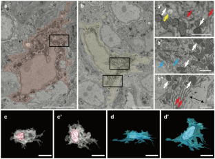
We previously clarified the histological characteristics of macrophages in the rat small intestine using serial block-face scanning electron microscopy (SBF-SEM). However, the regional differences in the characteristics of macrophages throughout the large intestine remain unknown. Here, we performed a pilot study to explore the regional differences in the ultrastructure of mucosal macrophages in the large intestine by using SBF-SEM analysis. SBF-SEM analysis conducted on the luminal side of the cecum and descending colon revealed macrophages as amorphous cells possessing abundant lysosomes and vacuoles. Macrophages in the cecum exhibited a higher abundance of lysosomes and a lower abundance of vacuoles than those in the descending colon. Macrophages with many intraepithelial cellular processes were observed beneath the intestinal superficial epithelium in the descending colon. Moreover, macrophages in contact with nerve fibers were more prevalent in the cecum than in the descending colon, and a subset of them surrounded a nerve bundle only in the cecum. In conclusion, the present pilot study suggested that the quantity of some organelles (lysosomes and vacuoles) in macrophages differed between the cecum and the descending colon and that there were some region-specific subsets of macrophages like nerve-associated macrophages in the cecum.
期刊介绍:
The journal publishes regular articles and reviews in the areas of molecular, cell, and supracellular biology. In particular, the journal intends to provide a forum for publishing data that analyze the supracellular, integrative actions of gene products and their impact on the formation of tissue structure and function. Submission of papers with an emphasis on structure-function relationships as revealed by recombinant molecular technologies is especially encouraged. Areas of research with a long-standing tradition of publishing in Cell & Tissue Research include:
- neurobiology
- neuroendocrinology
- endocrinology
- reproductive biology
- skeletal and immune systems
- development
- stem cells
- muscle biology.
 求助内容:
求助内容: 应助结果提醒方式:


