{"title":"ROCK1 deficiency preserves caveolar compartmentalization of signaling molecules and cell membrane integrity","authors":"Jianjian Shi, Lei Wei","doi":"10.1096/fba.2024-00015","DOIUrl":null,"url":null,"abstract":"<p>In this study, we investigated the roles of ROCK1 in regulating structural and functional features of caveolae located at the cell membrane of cardiomyocytes, adipocytes, and mouse embryonic fibroblasts (MEFs) as well as related physiopathological effects. Caveolae are small bulb-shaped cell membrane invaginations, and their roles have been associated with disease conditions. One of the unique features of caveolae is that they are physically linked to the actin cytoskeleton that is well known to be regulated by RhoA/ROCKs pathway. In cardiomyocytes, we observed that ROCK1 deficiency is coincident with an increased caveolar density, clusters, and caveolar proteins including caveolin-1 and -3. In the mouse cardiomyopathy model with transgenic overexpressing Gαq in myocardium, we demonstrated the reduced caveolar density at cell membrane and reduced caveolar protein contents. Interestingly, coexisting ROCK1 deficiency in cardiomyocytes can rescue these defects and preserve caveolar compartmentalization of β-adrenergic signaling molecules including β1-adrenergic receptor and type V/VI adenylyl cyclase. In cardiomyocytes and adipocytes, we detected that ROCK1 deficiency increased insulin signaling with increased insulin receptor activation in caveolae. In MEFs, we identified that ROCK1 deficiency increased caveolar and total levels of caveolin-1 and cell membrane repair ability after mechanical or chemical disruptions. Together, these results demonstrate that ROCK1 can regulate caveolae plasticity and multiple functions including compartmentalization of signaling molecules and cell membrane repair following membrane disruption by mechanical force and oxidative damage. These findings provide possible molecular insights into the beneficial effects of ROCK1 deletion/inhibition in cardiomyocytes, adipocytes, and MEFs under certain diseased conditions.</p>","PeriodicalId":12093,"journal":{"name":"FASEB bioAdvances","volume":"6 3","pages":"85-102"},"PeriodicalIF":2.5000,"publicationDate":"2024-02-23","publicationTypes":"Journal Article","fieldsOfStudy":null,"isOpenAccess":false,"openAccessPdf":"https://onlinelibrary.wiley.com/doi/epdf/10.1096/fba.2024-00015","citationCount":"0","resultStr":null,"platform":"Semanticscholar","paperid":null,"PeriodicalName":"FASEB bioAdvances","FirstCategoryId":"1085","ListUrlMain":"https://onlinelibrary.wiley.com/doi/10.1096/fba.2024-00015","RegionNum":0,"RegionCategory":null,"ArticlePicture":[],"TitleCN":null,"AbstractTextCN":null,"PMCID":null,"EPubDate":"","PubModel":"","JCR":"Q3","JCRName":"BIOCHEMISTRY & MOLECULAR BIOLOGY","Score":null,"Total":0}
引用次数: 0
Abstract
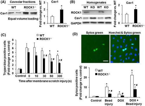
In this study, we investigated the roles of ROCK1 in regulating structural and functional features of caveolae located at the cell membrane of cardiomyocytes, adipocytes, and mouse embryonic fibroblasts (MEFs) as well as related physiopathological effects. Caveolae are small bulb-shaped cell membrane invaginations, and their roles have been associated with disease conditions. One of the unique features of caveolae is that they are physically linked to the actin cytoskeleton that is well known to be regulated by RhoA/ROCKs pathway. In cardiomyocytes, we observed that ROCK1 deficiency is coincident with an increased caveolar density, clusters, and caveolar proteins including caveolin-1 and -3. In the mouse cardiomyopathy model with transgenic overexpressing Gαq in myocardium, we demonstrated the reduced caveolar density at cell membrane and reduced caveolar protein contents. Interestingly, coexisting ROCK1 deficiency in cardiomyocytes can rescue these defects and preserve caveolar compartmentalization of β-adrenergic signaling molecules including β1-adrenergic receptor and type V/VI adenylyl cyclase. In cardiomyocytes and adipocytes, we detected that ROCK1 deficiency increased insulin signaling with increased insulin receptor activation in caveolae. In MEFs, we identified that ROCK1 deficiency increased caveolar and total levels of caveolin-1 and cell membrane repair ability after mechanical or chemical disruptions. Together, these results demonstrate that ROCK1 can regulate caveolae plasticity and multiple functions including compartmentalization of signaling molecules and cell membrane repair following membrane disruption by mechanical force and oxidative damage. These findings provide possible molecular insights into the beneficial effects of ROCK1 deletion/inhibition in cardiomyocytes, adipocytes, and MEFs under certain diseased conditions.

 求助内容:
求助内容: 应助结果提醒方式:


