Self-assembled peptide-dye nanostructures for in vivo tumor imaging and photodynamic toxicity

Raina M. Borum, Maurice Retout, Matthew N. Creyer, Yu-Ci Chang, Karlo Gregorio, Jesse V. Jokerst
{"title":"Self-assembled peptide-dye nanostructures for in vivo tumor imaging and photodynamic toxicity","authors":"Raina M. Borum, Maurice Retout, Matthew N. Creyer, Yu-Ci Chang, Karlo Gregorio, Jesse V. Jokerst","doi":"10.1038/s44303-024-00008-4","DOIUrl":null,"url":null,"abstract":"We report noncovalent assemblies of iRGD peptides and methylene blue dyes via electrostatic and hydrophobic stacking. These resulting nanomaterials could bind to cancer cells, image them with photoacoustic signal, and then treat them via photodynamic therapy. We first assessed the optical properties and physical properties of the materials. We then evaluated their utility for live cell targeting, in vivo imaging, and in vivo photodynamic toxicity. We tuned the performance of iRGD by adding aspartic acid (DD) or tryptophan doublets (WW) to the peptide to promote electrostatic or hydrophobic stacking with methylene blue, respectively. The iRGD-DD led to 150-nm branched nanoparticles, but iRGD-WW produced 200-nm nano spheres. The branched particles had an absorbance peak that was redshifted to 720 nm suitable for photoacoustic signal. The nanospheres had a peak at 680 nm similar to monomeric methylene blue. Upon continuous irradiation, the nanospheres and branched nanoparticles led to a 116.62% and 94.82% increase in reactive oxygen species in SKOV-3 cells relative to free methylene blue at isomolar concentrations suggesting photodynamic toxicity. Targeted uptake was validated via competitive inhibition. Finally, we used in vivo bioluminescent signal to monitor tumor burden and the effect of for photodynamic therapy: The nanospheres had little impact versus controls (p = 0.089), but the branched nanoparticles slowed SKOV-3 tumor burden by 75.9% (p < 0.05).","PeriodicalId":501709,"journal":{"name":"npj Imaging","volume":" ","pages":"1-9"},"PeriodicalIF":0.0000,"publicationDate":"2024-03-04","publicationTypes":"Journal Article","fieldsOfStudy":null,"isOpenAccess":false,"openAccessPdf":"https://www.nature.com/articles/s44303-024-00008-4.pdf","citationCount":"0","resultStr":null,"platform":"Semanticscholar","paperid":null,"PeriodicalName":"npj Imaging","FirstCategoryId":"1085","ListUrlMain":"https://www.nature.com/articles/s44303-024-00008-4","RegionNum":0,"RegionCategory":null,"ArticlePicture":[],"TitleCN":null,"AbstractTextCN":null,"PMCID":null,"EPubDate":"","PubModel":"","JCR":"","JCRName":"","Score":null,"Total":0}
引用次数: 0

Abstract

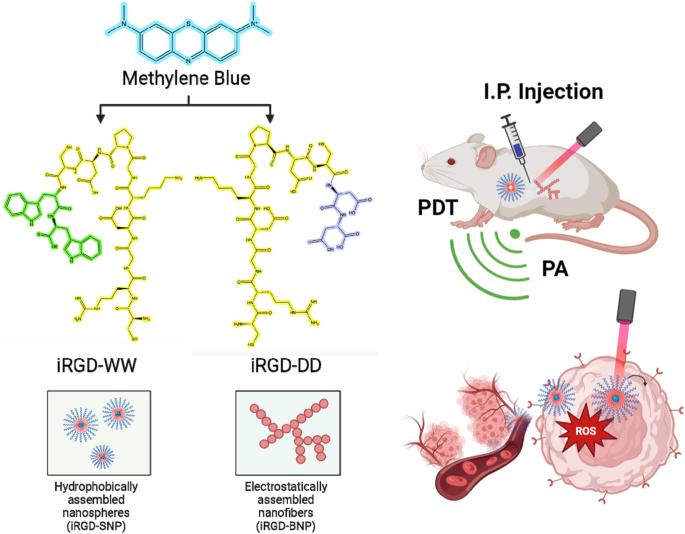
We report noncovalent assemblies of iRGD peptides and methylene blue dyes via electrostatic and hydrophobic stacking. These resulting nanomaterials could bind to cancer cells, image them with photoacoustic signal, and then treat them via photodynamic therapy. We first assessed the optical properties and physical properties of the materials. We then evaluated their utility for live cell targeting, in vivo imaging, and in vivo photodynamic toxicity. We tuned the performance of iRGD by adding aspartic acid (DD) or tryptophan doublets (WW) to the peptide to promote electrostatic or hydrophobic stacking with methylene blue, respectively. The iRGD-DD led to 150-nm branched nanoparticles, but iRGD-WW produced 200-nm nano spheres. The branched particles had an absorbance peak that was redshifted to 720 nm suitable for photoacoustic signal. The nanospheres had a peak at 680 nm similar to monomeric methylene blue. Upon continuous irradiation, the nanospheres and branched nanoparticles led to a 116.62% and 94.82% increase in reactive oxygen species in SKOV-3 cells relative to free methylene blue at isomolar concentrations suggesting photodynamic toxicity. Targeted uptake was validated via competitive inhibition. Finally, we used in vivo bioluminescent signal to monitor tumor burden and the effect of for photodynamic therapy: The nanospheres had little impact versus controls (p = 0.089), but the branched nanoparticles slowed SKOV-3 tumor burden by 75.9% (p < 0.05).
Abstract Image

用于体内肿瘤成像和光动力毒性的自组装肽染料纳米结构
我们报告了 iRGD 肽和亚甲基蓝染料通过静电和疏水堆积的非共价组装。这些纳米材料可与癌细胞结合,利用光声信号对其成像,然后通过光动力疗法对其进行治疗。我们首先评估了这些材料的光学特性和物理特性。然后,我们评估了它们在活细胞靶向、体内成像和体内光动力毒性方面的实用性。我们通过在肽中添加天冬氨酸(DD)或色氨酸双酯(WW)来调节 iRGD 的性能,以分别促进与亚甲基蓝的静电或疏水堆叠。iRGD-DD 产生了 150 纳米的支化纳米颗粒,而 iRGD-WW 则产生了 200 纳米的纳米球。支化颗粒的吸光度峰红移到 720 纳米,适合光声信号。纳米球在 680 纳米处有一个与单体亚甲基蓝相似的峰值。连续照射时,纳米球和支化纳米粒子导致 SKOV-3 细胞中的活性氧相对于等摩尔浓度的游离亚甲基蓝分别增加了 116.62% 和 94.82%,这表明它们具有光动力毒性。通过竞争性抑制验证了靶向吸收。最后,我们利用体内生物发光信号来监测肿瘤负荷和光动力疗法的效果:与对照组相比,纳米球几乎没有影响(p = 0.089),但支化纳米粒子使 SKOV-3 肿瘤负荷减少了 75.9%(p < 0.05)。
本文章由计算机程序翻译,如有差异,请以英文原文为准。
求助全文
约1分钟内获得全文 求助全文
来源期刊
自引率
0.00%
发文量
0
×
引用
GB/T 7714-2015
复制
MLA
复制
APA
复制
导出至
BibTeX EndNote RefMan NoteFirst NoteExpress
×
提示
您的信息不完整,为了账户安全,请先补充。
现在去补充
×
提示
您因"违规操作"
具体请查看互助需知
我知道了
×
提示
确定
请完成安全验证×
copy
已复制链接
快去分享给好友吧!
我知道了
右上角分享
点击右上角分享
0
联系我们:info@booksci.cn Book学术提供免费学术资源搜索服务,方便国内外学者检索中英文文献。致力于提供最便捷和优质的服务体验。 Copyright © 2023 布克学术 All rights reserved.
京ICP备2023020795号-1
ghs 京公网安备 11010802042870号
Book学术文献互助
Book学术文献互助群
群 号:481959085
Book学术官方微信