Cellular and molecular control of vertebrate somitogenesis
IF 81.3
1区 生物学
Q1 CELL BIOLOGY
引用次数: 0
Abstract
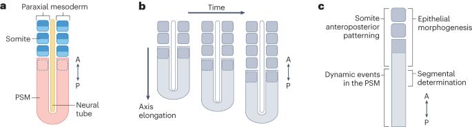
Segmentation is a fundamental feature of the vertebrate body plan. This metameric organization is first implemented by somitogenesis in the early embryo, when paired epithelial blocks called somites are rhythmically formed to flank the neural tube. Recent advances in in vitro models have offered new opportunities to elucidate the mechanisms that underlie somitogenesis. Notably, models derived from human pluripotent stem cells introduced an efficient proxy for studying this process during human development. In this Review, we summarize the current understanding of somitogenesis gained from both in vivo studies and in vitro studies. We deconstruct the spatiotemporal dynamics of somitogenesis into four distinct modules: dynamic events in the presomitic mesoderm, segmental determination, somite anteroposterior polarity patterning, and epithelial morphogenesis. We first focus on the segmentation clock, as well as signalling and metabolic gradients along the tissue, before discussing the clock and wavefront and other models that account for segmental determination. We then detail the molecular and cellular mechanisms of anteroposterior polarity patterning and somite epithelialization. Somite formation, crucial for organization of the segmental pattern of vertebrate embryos, depends on the oscillatory expression of segmentation clock genes. Novel in vitro models of somitogenesis have provided insights into the spatiotemporal dynamics of gene expression, signalling and metabolic gradients that enable somite formation and patterning.
脊椎动物体节发生的细胞和分子控制。
分割是脊椎动物身体结构的一个基本特征。在早期胚胎中,体节发生首先实现了这种元组织,此时称为体节的成对上皮块有节奏地形成,位于神经管两侧。体外模型的最新进展为阐明体节发生的机制提供了新的机会。值得注意的是,源自人类多能干细胞的模型为研究人类发育过程中的这一过程提供了有效的替代方法。在本综述中,我们总结了目前从体内研究和体外研究中获得的对体节发生的理解。我们将体细胞发生的时空动态解构为四个不同的模块:前绒毛中胚层的动态事件、节段决定、体细胞前后极性模式化和上皮形态发生。在讨论时钟和波前以及其他解释节段决定的模型之前,我们首先关注节段时钟以及沿组织的信号和代谢梯度。然后,我们将详细介绍前胸极性模式化和体节上皮化的分子和细胞机制。
本文章由计算机程序翻译,如有差异,请以英文原文为准。
求助全文
约1分钟内获得全文
求助全文
来源期刊
CiteScore
173.60
自引率
0.50%
发文量
118
审稿时长
6-12 weeks
期刊介绍:
Nature Reviews Molecular Cell Biology is a prestigious journal that aims to be the primary source of reviews and commentaries for the scientific communities it serves. The journal strives to publish articles that are authoritative, accessible, and enriched with easily understandable figures, tables, and other display items. The goal is to provide an unparalleled service to authors, referees, and readers, and the journal works diligently to maximize the usefulness and impact of each article. Nature Reviews Molecular Cell Biology publishes a variety of article types, including Reviews, Perspectives, Comments, and Research Highlights, all of which are relevant to molecular and cell biologists. The journal's broad scope ensures that the articles it publishes reach the widest possible audience.
文献相关原料
| 公司名称 | 产品信息 | 采购帮参考价格 |
|---|
 求助内容:
求助内容: 应助结果提醒方式:


