Two Rare Benign Lesions on 18F-FDG PET/CT: Peliosis Hepatis and SANT.
IF 1.1
Q4 RADIOLOGY, NUCLEAR MEDICINE & MEDICAL IMAGING
Molecular Imaging and Radionuclide Therapy
Pub Date : 2024-02-22
DOI:10.4274/mirt.galenos.2023.02328
引用次数: 0
Abstract
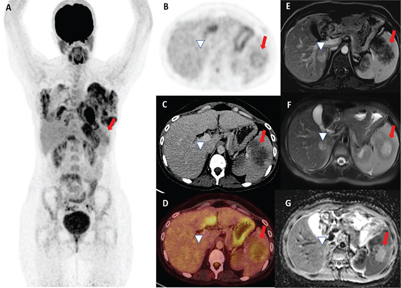
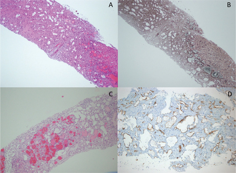
Peliosis hepatis (PH) and sclerosing angiomatoid nodular transformation of the spleen are uncommon benign lesions. Diagnosis can be difficult in some patients. Herein, we present the case of a 28-year-old woman referred with abdominal pain who had spleen lesions. 18F-fluorodeoxyglucose (FDG) positron emission tomography/computed tomography revealed multiple non-FDG avid lesions in the liver and hypermetabolic lesions in the spleen. In addition, abdominal magnetic resonance imaging was performed. Histopathology revealed sclerosing angiomatoid nodular transformation in the spleen and PH in the liver.
18F-FDG PET/CT 上的两种罕见良性病变:肝脓肿和 SANT。
肝脓肿(PH)和脾硬化性血管瘤样结节变是不常见的良性病变。有些患者很难确诊。在此,我们介绍了一例因腹痛而转诊的 28 岁女性脾脏病变病例。18F- 氟脱氧葡萄糖(FDG)正电子发射断层扫描/计算机断层扫描显示肝脏有多个非 FDG 阳性病变,脾脏有高代谢病变。此外,还进行了腹部磁共振成像检查。组织病理学显示,脾脏出现硬化性血管瘤样结节转化,肝脏出现 PH。
本文章由计算机程序翻译,如有差异,请以英文原文为准。
求助全文
约1分钟内获得全文
求助全文
来源期刊

Molecular Imaging and Radionuclide Therapy
RADIOLOGY, NUCLEAR MEDICINE & MEDICAL IMAGING-
CiteScore
1.30
自引率
0.00%
发文量
50
期刊介绍:
Molecular Imaging and Radionuclide Therapy (Mol Imaging Radionucl Ther, MIRT) is publishes original research articles, invited reviews, editorials, short communications, letters, consensus statements, guidelines and case reports with a literature review on the topic, in the field of molecular imaging, multimodality imaging, nuclear medicine, radionuclide therapy, radiopharmacy, medical physics, dosimetry and radiobiology.
 求助内容:
求助内容: 应助结果提醒方式:


